Every living organism, from microscopic bacteria to giant trees and animals, is made up of one or
more cells. A cell is the smallest structural and functional unit of life capable of performing all
essential life processes such as nutrition, respiration, excretion, growth and reproduction.
Just as bricks are the building blocks of a building, cells are the building blocks of all living
organisms. The study of cells helps us understand how living organisms function and how diseases
develop and can be treated.
📘 Definition
Cell: A cell is the basic structural, functional and biological unit of life.
It is the smallest unit capable of carrying out all life activities independently.
In simple words, a cell can be defined as:
"The smallest living unit that can exist independently and perform all essential functions of life."
🤔 Did You Know?
Why are Cells Called the Fundamental Unit of Life?
All living organisms are made up of cells.
Cells carry out all metabolic activities of organisms.
Growth occurs because of an increase in the number of cells.
Repair of damaged tissues takes place through cell division.
Reproduction in many organisms occurs through cells.
All hereditary information is ultimately stored inside cells.
Therefore, no living organism can exist without cells, making them the
fundamental unit of life.
🏛️ Historical Note
Historical Background of Cell Discovery
Scientist
Contribution
Year
Robert Hooke
Observed thin slices of cork and coined the term "cell".
1665
Antonie van Leeuwenhoek
Observed living cells for the first time.
1674
Robert Brown
Discovered the nucleus.
1831
Matthias Schleiden
Proposed that all plants are made of cells.
1838
Theodor Schwann
Proposed that all animals are made of cells.
1839
Rudolf Virchow
Proposed that new cells arise from pre-existing cells.
1855
📌 Cell Theory
📘
Definition
Cell Theory was proposed by Schleiden and Schwann and later modified by
Rudolf Virchow.
Postulates of Cell Theory
Helps in understanding growth and development of organisms.
Provides knowledge about heredity and genetics.
Explains causes of many diseases such as cancer and infections.
Forms the basis of biotechnology and genetic engineering.
Helps in medical research and development of medicines.
Useful in tissue culture and regenerative medicine.
🔷 General Characteristics of Cells
🔷Characteristics
Cells are microscopic in most organisms.
Cells vary greatly in size and shape.
Every cell is surrounded by a plasma membrane.
Cells contain living material called protoplasm.
Cells possess genetic material in the form of DNA.
Cells can perform metabolism and produce energy.
Cells can grow and divide.
🗂️ Types / Category
Main Parts of a Typical Cell
Plasma Membrane (Cell Membrane)
The outermost, selectively permeable covering of the cell that regulates the entry and exit of substances, maintaining the cell's internal environment.
Cytoplasm
The fluid region inside the plasma membrane that houses the cell organelles and serves as the site for various metabolic chemical reactions.
Nucleus
The double-membrane-bound control center of the cell containing genetic material (chromosomes/DNA) that directs growth, reproduction, and cellular activities.
📌
Note
Various specialized structures called cell organelles are suspended in the
cytoplasm and perform specific functions.
📌 What are Cell Organelles?
Cell organelles are specialized structures present inside cells that perform specific functions
necessary for survival of the cell.
Examples:
Nucleus
Mitochondria
Endoplasmic Reticulum
Golgi Apparatus
Lysosomes
Plastids
Vacuoles
Ribosomes
ℹ️ Cell as a Miniature Factory
Cell Part
Acts Like
Function
Nucleus
Manager's Office
Controls activities of the cell
Plasma Membrane
Security Gate
Regulates entry and exit of materials
Mitochondria
Power House
Produces energy
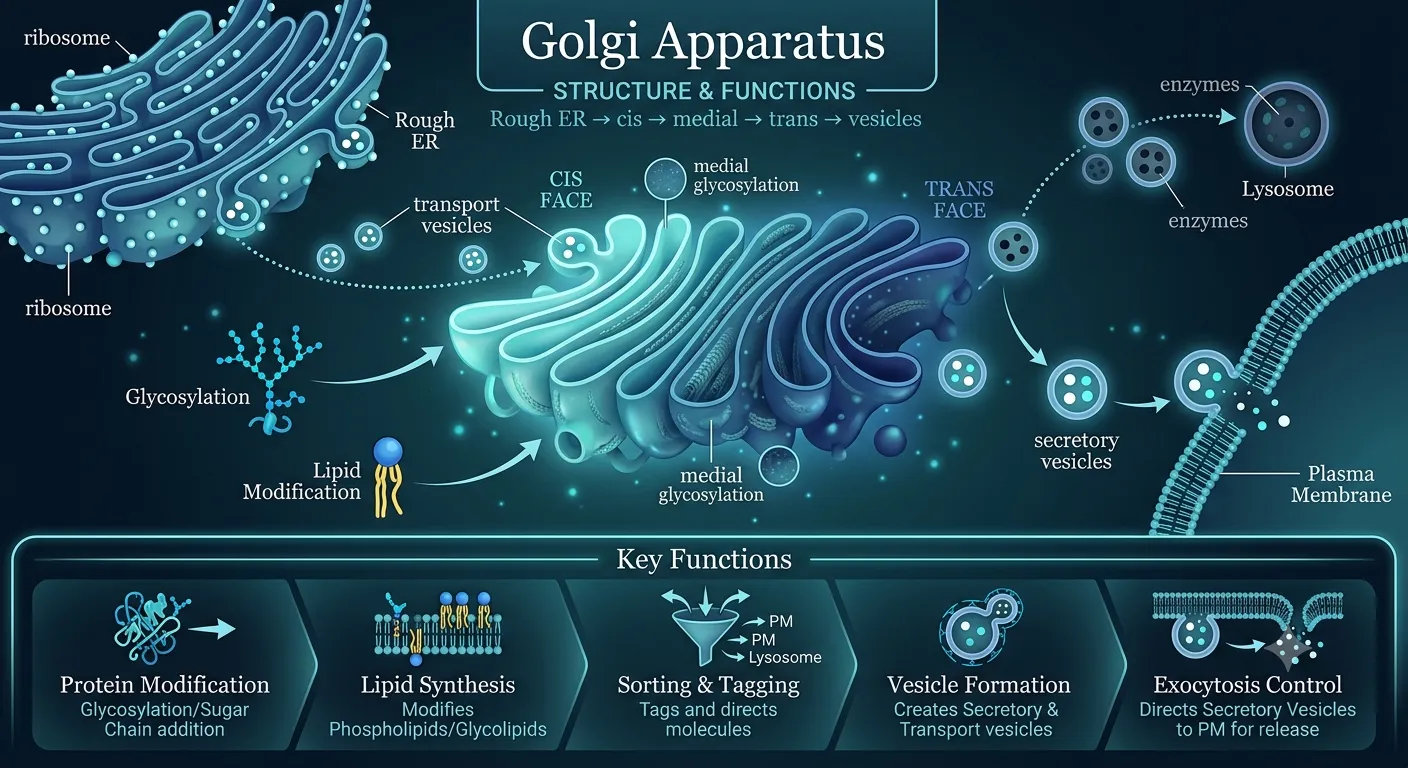
Golgi Apparatus
Packing Department
Packaging and secretion
Endoplasmic Reticulum
Transport Network
Transportation of materials
Lysosomes
Cleaning Department
Digestion and waste removal
✏️ Examples for Better Understanding
Example 1: Amoeba is made of only one cell, yet it can move, digest food and reproduce.
Example 2: Human beings have trillions of cells organized into tissues, organs and organ systems.
Example 3: A nerve cell can be more than one metre long, whereas a red blood cell is only about 7 micrometres in diameter.
✏️ Solved Example
Why is a cell called both the structural and functional unit of life?
Structural because all organisms are built from cells.
Functional because all life processes occur within cells.
Organisms → Made of Cells → Cells Perform Life Processes → Therefore Cells are Structural and Functional Units.
A cell is called the structural unit because all organisms are made of cells, and it is called
the functional unit because every life process such as nutrition, respiration and reproduction
takes place within cells.
🌟 Points Important for CBSE Board Examination
Definition of cell is frequently asked in one-mark questions.
Contributions of Hooke, Leeuwenhoek, Schleiden, Schwann and Virchow are important.
Postulates of Cell Theory are repeatedly asked in examinations.
Difference between structural and functional unit of life is important.
Functions of plasma membrane, nucleus and cytoplasm should be remembered.
Living organisms exhibit a definite structural organisation. In multicellular organisms, millions of cells
work together in a coordinated manner to perform life processes. The organisation occurs in a hierarchical
manner:
Cell → Tissue → Organ → Organ System → Organism
The cell is the fundamental structural and functional unit from which higher levels of organisation arise.
Each cell is enclosed by a protective boundary called the plasma membrane, which separates
the living contents of the cell from the surrounding environment and regulates the movement of substances.
📘 Definition
Plasma Membrane (Cell Membrane)
The plasma membrane, also called the cell membrane, is the thin,
living, flexible outer boundary of the cell that surrounds the cytoplasm and separates the internal
contents of the cell from the external environment.
"The plasma membrane is a selectively permeable membrane that regulates the movement of substances
into and out of the cell."
📌 Location of Plasma Membrane
In animal cells, it forms the outermost boundary of the cell.
In plant cells, it lies immediately inside the cell wall.
It surrounds the cytoplasm and encloses all cell organelles.
ℹ️ Chemical Composition of Plasma Membrane
The plasma membrane is mainly composed of:
Phospholipids
Proteins
Small quantities of carbohydrates and cholesterol (in animal cells)
Because of this arrangement, phospholipids automatically form a
phospholipid bilayer, providing both stability and flexibility to the membrane.
💡 Fluid Mosaic Concept (Advanced Concept)
According to the Fluid Mosaic Model, the plasma membrane behaves like a
fluid sheet of phospholipids in which proteins are embedded.
The membrane is not rigid; instead, lipid and protein molecules can move sideways,
making it flexible and dynamic.
This flexibility allows:
Movement of materials across the membrane.
Growth and repair of cells.
Formation of vesicles during endocytosis and exocytosis.
Changes in cell shape.
Although the detailed Fluid Mosaic Model is studied in higher classes, understanding membrane
flexibility helps explain many cellular processes in Class IX.
🏷️ Properties of Plasma Membrane
Properties
Structure
Extremely thin, delicate, and visible only under an electron microscope.
Vitality
Living, dynamic, and metabolically active outer boundary of the cell.
Flexibility
Highly flexible and elastic, allowing the cell to engulf food and other substances (endocytosis).
Permeability
Selectively permeable, permitting the entry and exit of only select substances.
Regeneration
Capable of self-repair and automatically sealing minor ruptures.
Fluidity
Exhibits fluid characteristics due to its lipid bilayer and embedded proteins.
🤔 Did You Know?
Why is Plasma Membrane Called Selectively Permeable?
A selectively permeable membrane allows certain substances to pass through it while restricting
the movement of others.
Substance
Movement Through Membrane
Oxygen (O₂)
Passes freely
Carbon dioxide (CO₂)
Passes freely
Water
Passes under specific conditions
Large molecules
Usually require special transport mechanisms
This selective permeability helps maintain the proper internal environment of the cell,
a condition called cellular homeostasis.
🌟 Functions of Plasma Membrane
Forms the protective outer covering of the cell.
Separates cell contents from the external environment.
Controls entry and exit of substances.
Maintains the chemical composition of the cytoplasm.
Molecules are continuously moving due to their kinetic energy. As a result, substances move from
regions of higher concentration to regions of lower concentration until uniform distribution is achieved.
"Diffusion is the spontaneous movement of particles from a region of higher concentration to a region
of lower concentration."
✏️ Examples of Diffusion
The smell of perfume spreading throughout a room.
The aroma of food reaching us from the kitchen.
Carbon dioxide moving out of cells.
Oxygen entering cells during respiration.
🌟 Significance of Diffusion in Cells
Provides oxygen to cells.
Removes carbon dioxide produced during respiration.
Facilitates exchange of gases between cells and surroundings.
✏️ Example: Diffusion During Breathing
The concentration of oxygen is generally higher in the air present inside the lungs than in blood.
Therefore, oxygen diffuses from the lungs into the blood.
Similarly, carbon dioxide concentration is higher in blood than in lung air.
Therefore, carbon dioxide diffuses from blood into the lungs and is exhaled.
🌟 Factors Affecting Diffusion
Difference in concentration
Temperature
Surface area available for diffusion
Size of particles
Nature of the membrane
✏️ Example
Concept Builder
Why can oxygen and carbon dioxide move through the plasma membrane without using energy?
Both gases are small molecules.
They move from higher concentration to lower concentration.
This movement occurs by diffusion.
No cellular energy is required.
Oxygen and carbon dioxide are small molecules that move through the plasma membrane by diffusion,
that is, from regions of higher concentration to lower concentration. Since diffusion is a passive
process, no energy is required.
🔗 Real-Life Analogy
The plasma membrane behaves like a security gate of a school. Students and teachers may enter or leave
according to certain rules, while unauthorized persons are restricted. Similarly, the plasma membrane
selectively regulates the movement of substances.
📋 CBSE Case Study (HOTS)<
A biology student placed a few living cells in a chamber containing high oxygen concentration.
After some time, oxygen molecules were found inside the cells.
Questions
Which property of the plasma membrane is demonstrated?
By which process did oxygen enter the cells?
Was cellular energy required?
Answers
Selectively permeable nature of the plasma membrane.
Diffusion.
No. Diffusion is a passive process.
⚡ Exam Tip
Always write "selectively permeable membrane", not semipermeable membrane.
Remember that diffusion occurs from higher concentration to lower concentration.
Do not write that the membrane is made only of proteins.
State that the membrane consists mainly of phospholipids and proteins.
Diffusion does not require energy.
❌ Common Mistakes
Confusing diffusion with osmosis.
Writing that all substances can freely cross the plasma membrane.
Forgetting that the plasma membrane is flexible and living.
Stating that diffusion occurs from lower concentration to higher concentration.
Living organisms are highly organised systems. In multicellular organisms, individual cells do not work
independently. Similar cells group together to form tissues, tissues combine to form organs, organs work
together as organ systems, and all organ systems together constitute an organism.
This arrangement is called the levels of structural organisation.
Cell → Tissue → Organ → Organ System → Organism
📌 Levels of Organisation
Level
Definition
Examples
Cell
Smallest structural and functional unit of life.
Nerve cell, muscle cell, red blood cell
Tissue
Group of similar cells performing a common function.
Muscular tissue, xylem tissue
Organ
Group of different tissues performing a specific function.
Heart, stomach, leaf, root
Organ System
Group of organs working together.
Digestive system, respiratory system
Organism
Entire living individual formed by all organ systems.
Human being, mango tree
🤔 Did You Know?
Why is Structural Organisation Important?
Ensures division of labour among cells and organs.
Increases efficiency of biological functions.
Allows specialised functions to be performed simultaneously.
Maintains coordination and homeostasis in the body.
Makes complex life processes possible.
✏️ Example
Human Body:
Muscle Cell → Muscular Tissue → Heart → Circulatory System → Human Being
The plasma membrane regulates the movement of substances between the cell and its surroundings.
This movement is known as transport across the plasma membrane.
🗂️ Types / Category
Transport mechanismsPassive TransportActive TransportConcentration Gradient
Passive Transport
Definition Passive transport is the movement of molecules or ions across the plasma membrane
from a region of higher concentration to a region of lower concentration without the expenditure
of cellular energy. Characteristics
No energy is required.
Movement occurs along the concentration gradient.
Generally involves movement from higher concentration to lower concentration.
Occurs spontaneously.
Examples of Passive Transport
Diffusion of oxygen into cells.
Diffusion of carbon dioxide out of cells.
Movement of water by osmosis.
Active Transport
Definition
Active transport is the movement of molecules or ions across a membrane from a region
of lower concentration to a region of higher concentration against the concentration gradient,
requiring expenditure of cellular energy.
Characteristics
Requires cellular energy in the form of ATP.
Occurs against the concentration gradient.
Usually assisted by transport proteins present in the membrane.
Highly selective and regulated.
Essential for maintaining the chemical composition of cells.
Biological Importance
Absorption of mineral ions by plant roots.
Absorption of glucose and amino acids in the small intestine.
Maintenance of ionic balance in nerve cells.
Transport of nutrients against concentration differences.
What is Concentration Gradient?
📌
Note
The difference in concentration of a substance between two regions is called the
concentration gradient.
Molecules naturally tend to move from regions of higher concentration toward regions of lower
concentration until equilibrium is established.
⚖️ Difference Between Active and Passive Transport
Basis of Comparison
Passive Transport
Active Transport
Energy Requirement
No energy required
Requires ATP energy
Direction of Movement
High concentration → Low concentration
Low concentration → High concentration
Concentration Gradient
Along the concentration gradient
Against the concentration gradient
Transport Proteins
May or may not be involved
Usually required
Examples
Diffusion, osmosis
Mineral uptake by roots, ion pumps
✏️ Example
Concept Builder
Why can oxygen enter cells without energy expenditure, whereas mineral ions often require energy for absorption?
Oxygen generally moves from higher concentration to lower concentration.
This movement occurs by diffusion.
Mineral ions may need to move from lower concentration outside the cell to higher concentration inside the cell.
This movement occurs against the concentration gradient and therefore requires energy.
Oxygen enters cells through passive transport by diffusion and does not require energy.
Mineral ions are frequently transported against the concentration gradient, so ATP energy is required,
resulting in active transport.
📋 CBSE Competency-Based Case Study (HOTS)
Plant roots were placed in a dilute mineral solution. Scientists observed that certain mineral ions
accumulated inside root cells even though their concentration was already higher inside the cells than
outside.
Questions
Which type of transport is taking place?
Does this process require energy?
Why cannot this process occur by diffusion?
Answers
Active transport.
Yes. ATP energy is required.
Diffusion always occurs from higher concentration to lower concentration, whereas here ions move in the opposite direction.
⚡ Exam Tip
Remember: Passive transport is a downhill movement of molecules; active transport is an uphill movement.
Diffusion and osmosis are examples of passive transport.
Active transport always requires ATP energy.
Plant roots absorbing minerals is a favourite CBSE application-based question.
Concentration gradient-based diagrams are frequently asked in competency-based assessments.
❌ Common Mistakes
Writing that active transport occurs from higher concentration to lower concentration.
Writing that diffusion requires energy.
Confusing diffusion with active transport.
Ignoring the role of ATP in active transport.
Writing that all transport processes require membrane proteins.
Water molecules are continuously moving and also follow the principle of diffusion.
However, when the movement of water occurs through a
selectively permeable membrane, the process is called
osmosis.
📘 Definition
Osmosis is the movement (diffusion) of water molecules through a selectively permeable membrane
from a region of higher water concentration (lower solute concentration) to a region of lower
water concentration (higher solute concentration).
In simpler words, water moves from a dilute solution to a concentrated solution through a selectively permeable membrane.
Net Diffusion of Water
Since dissolved substances occupy space between water molecules, the concentration of water
decreases as the amount of dissolved solute increases.
Therefore, water always moves:
Higher Water Concentration → Lower Water Concentration
Water molecules possess kinetic energy and remain in continuous random motion.
If two solutions of different concentrations are separated by a selectively permeable membrane,
water molecules move from the region containing more water molecules to the region containing
fewer water molecules until equilibrium is established.
Conditions Necessary for Osmosis
A selectively permeable membrane must be present.
The two solutions must have different concentrations.
Water molecules must be able to move through the membrane.
🤔 What is a Selectively Permeable Membrane?
A selectively permeable membrane allows certain molecules, especially water molecules,
to pass through it while restricting the movement of many dissolved substances.
The plasma membrane of the cell behaves as a selectively permeable membrane and
therefore permits osmosis to occur.
📎 Role of Solute Concentration
The movement of water across the plasma membrane depends upon the amount of dissolved
substances present in water.
Solution
Solute Concentration
Water Concentration
Dilute Solution
Low
High
Concentrated Solution
High
Low
Hence, water moves from the dilute solution toward the concentrated solution.
🔷 Characteristics of Osmosis
🔷Characteristics
Osmosis is a type of passive transport.
No ATP energy is required.
Only solvent molecules (usually water) move.
Movement occurs through a selectively permeable membrane.
Water moves from lower solute concentration to higher solute concentration.
Osmosis continues until equilibrium is established.
🗂️ Types / Category
Types of Solutions in Relation to Cells
Hypotonic Solution
Definition: A solution surrounding the cell that has a higher water concentration (lower solute concentration) than the cell sap. Explanation: Due to osmosis, water molecules pass through the cell membrane in both directions, but the net flow is into the cell. This causes the cell to swell, creating turgor pressure in plant cells or potentially bursting animal cells. Examples: Placing dried raisins in plain water (they swell up), or red blood cells placed in distilled water.
Hypertonic Solution
Definition: A solution surrounding the cell that has a lower water concentration (higher solute concentration) than the cell sap. Explanation: Since the outside solution is highly concentrated, water molecules spontaneously exit the cell via osmosis. The net outflow of water causes the cell to shrink. In plant cells, this causes the cell contents to shrink away from the cell wall (plasmolysis). Examples: Placing grapes or red blood cells in a highly concentrated salt or sugar solution (they shrink/crenate).
Isotonic Solution
Definition: A solution surrounding the cell that has exactly the same water and solute concentration as the cell sap. Explanation: Water molecules cross the cell membrane in both directions, but the rate of entry is equal to the rate of exit. Because there is no net movement of water, the cell maintains its original size and shape. Examples: Placing animal cells in a 0.9% physiological saline solution, where the cells remain stable.
🌟 Biological Significance of Osmosis
Helps root hairs absorb water from the soil.
Maintains turgidity and rigidity of plant cells.
Facilitates movement of water from one cell to another.
Maintains proper water balance in living organisms.
Helps in opening and closing of stomata.
Prevents excessive dehydration of cells.
✏️ Examples of Osmosis in Daily Life
Dry raisins swell when placed in water.
Wilted vegetables become fresh after immersion in water.
Cucumber slices lose water when salt is sprinkled on them.
Preservation of pickles using concentrated salt solution.
Roots absorb water from moist soil.
⚖️ Difference Between Diffusion and Osmosis
Basis
Diffusion
Osmosis
Substance Moving
Any molecules
Only water molecules
Membrane Requirement
Not essential
Selectively permeable membrane is essential
Direction
Higher concentration to lower concentration
Higher water concentration to lower water concentration
Energy Requirement
No energy required
No energy required
✏️ Example
Concept Builder
Why do dry raisins swell when placed in water?
The raisin contains concentrated sugar solution.
Water outside the raisin has higher water concentration.
The raisin skin acts as a selectively permeable membrane.
Water enters the raisin through osmosis.
Water outside (high water concentration)
→ Selectively permeable membrane
→ Water enters raisin
→ Raisin swells.
Dry raisins swell because water enters them through osmosis from the surrounding dilute solution
into the concentrated solution present inside the raisins.
📋 CBSE Competency-Based Case Study (HOTS)
A student placed two potato cups in separate beakers. One beaker contained pure water and the
other contained concentrated sugar solution. After some time, the potato cup placed in pure water
became firm and swollen, whereas the other became soft and shrivelled.
Questions
Which process is responsible for these observations?
Why did the potato cup in water become swollen?
Why did the potato cup in concentrated sugar solution shrink?
Answers
Osmosis.
Water entered the potato cells by osmosis.
Water moved out of the potato cells into the concentrated sugar solution.
⚡ Exam Tip
Remember: Osmosis is diffusion of water only.
Always mention "through a selectively permeable membrane" in the definition.
Water moves from dilute solution to concentrated solution.
Osmosis is a passive process and does not require ATP.
Questions involving raisins, potato cups and root hairs are frequently asked in CBSE examinations.
❌ Common Mistakes
Writing that osmosis is movement of solute molecules.
Forgetting to mention selectively permeable membrane in the definition.
Confusing osmosis with diffusion.
Writing that osmosis requires energy.
Writing that water moves from concentrated solution to dilute solution.
A hypotonic solution is a solution that has a lower solute concentration and a higher water concentration than the cell sap.
Since the surrounding solution contains more water molecules than the cell, water enters the cell
through osmosis.
Outside Solution: High Water Concentration, Low Solute Concentration Inside Cell: Lower Water Concentration, Higher Solute Concentration
Movement of Water
Water moves from the surrounding dilute solution into the cell through the selectively permeable
plasma membrane.
Outside of Cell → Inside of Cell
This inward movement of water is called endosmosis.
🤔 Did You Know?
What is Endosmosis?
Endosmosis is the movement of water into a cell through a selectively permeable membrane when the cell is placed in a hypotonic solution.
Endosmosis causes the volume of the cell to increase because water continuously enters the cell.
🗂️ Effect of Hypotonic Solution on
Plant CellsAnimal Cells
Plant Cells
When a plant cell is placed in a hypotonic solution, water enters the cell by osmosis.
Vacuole absorbs water and enlarges.
Cytoplasm expands.
The plasma membrane presses against the cell wall.
The cell becomes swollen and firm.
The plant cell does not burst because the rigid cell wall exerts an opposing force called
wall pressure.
The pressure exerted by the cell contents against the cell wall is called
turgor pressure.
A fully swollen plant cell is called a turgid cell.
Animal Cells
Animal cells lack a cell wall. Therefore, when water enters continuously through osmosis,
the cell swells considerably.
If excessive water enters the cell, the plasma membrane may rupture and the cell may burst.
The bursting of an animal cell due to excessive entry of water is called
cytolysis.
🗒️ Mechanism Of Hypotonic Solution
Step 1: Outside solution contains more water molecules. Step 2: Water diffuses through the selectively permeable membrane. Step 3: Water enters the cell by endosmosis. Step 4: Cell volume increases. Step 5: Plant cells become turgid, whereas animal cells may burst.
🌟 Biological Significance of Hypotonic Solutions
Maintains turgidity of plant cells.
Provides mechanical support to herbaceous plants.
Keeps leaves and stems erect.
Facilitates expansion and growth of young plant tissues.
Helps maintain normal cellular functions.
✏️ Examples of Hypotonic Solutions
Pure water surrounding a raisin.
Fresh water surrounding certain aquatic organisms.
Distilled water around plant cells.
Water absorbed by root hairs from moist soil.
🛠️ Application
Applications and Everyday Observations
Dry raisins swell when soaked in water.
Wilted vegetables become fresh after being placed in water.
Plant leaves become firm after watering.
Seeds absorb water and swell during germination.
✏️ Concept Builder
Why do wilted spinach leaves become crisp and firm when placed in water?
Water has a higher water concentration than the cell sap.
Water enters cells by osmosis.
Cells become turgid due to endosmosis.
Turgid cells make leaves firm and erect.
Water outside → Endosmosis → Cells become turgid → Leaves become firm.
Wilted spinach leaves become crisp because water enters their cells through endosmosis.
The cells regain turgidity, making the leaves firm and fresh.
📋 CBSE Competency-Based Case Study (HOTS)
A student placed a few raisins in a beaker containing pure water and left them overnight.
The next day, the raisins appeared swollen.
Questions
Which type of solution surrounded the raisins?
Name the process responsible for swelling.
What is the movement of water into the raisin called?
Answers
Hypotonic solution.
Osmosis.
Endosmosis.
🌟 Points Important for Board Examinations
Hypotonic solution has lower solute concentration and higher water concentration than the cell sap.
Water enters the cell through osmosis.
The inward movement of water is called endosmosis.
Plant cells become turgid but do not burst because of the cell wall.
Animal cells may burst in strongly hypotonic solutions.
❌ Common Mistakes
Writing that water moves out of the cell in a hypotonic solution.
Confusing hypotonic and hypertonic solutions.
Writing that plant cells burst in hypotonic solutions.
Forgetting the term endosmosis.
Writing that hypotonic solutions have higher solute concentration.
An isotonic solution is a solution that has the same solute concentration and the same water concentration as that of the cell sap.
Since the concentration of water molecules is equal inside and outside the cell, water molecules move
in both directions at the same rate. Therefore, there is
no net movement of water across the plasma membrane.
Movement of Water in an Isotonic Solution
Water entering the cell = Water leaving the cell
Although water molecules continuously move across the plasma membrane,
the amount of water entering the cell is exactly equal to the amount of water leaving it.
Therefore, the size, shape and volume of the cell remain unchanged.
Dynamic Equilibrium
The condition in which molecules continue to move but there is no overall change in concentration
is called dynamic equilibrium.
In an isotonic solution, the cell and the surrounding medium are in dynamic equilibrium.
🗂️ Effect of Hypotonic Solution on
Plant CellsAnimal Cell
Plant Cell
No net movement of water occurs.
The cell neither gains nor loses water.
The vacuole does not enlarge significantly.
The cell remains in its normal state.
The cell is neither fully turgid nor plasmolysed.
Animal Cell
No net movement of water occurs.
The cell maintains its normal size and shape.
The cell neither swells nor shrinks.
Normal physiological activities continue efficiently.
🗒️ Mechanism Of An Isotonic Solution
Step 1: Water concentration is equal inside and outside the cell. Step 2: Water molecules move continuously in both directions. Step 3: Equal amounts of water enter and leave the cell. Step 4: Cell volume remains constant.
🌟 Biological Significance of Isotonic Solutions
Maintains normal shape and size of cells.
Prevents excessive swelling or shrinkage of cells.
Ensures proper functioning of animal cells and tissues.
Helps maintain internal water balance in organisms.
Widely used in medicine for intravenous fluids and saline solutions.
✏️ Examples of Isotonic Solutions
Red blood cells placed in normal saline solution (0.9% sodium chloride solution).
Certain intravenous fluids administered in hospitals.
Body fluids surrounding many cells are nearly isotonic in nature.
🌟 Important Note for Competitive Examinations
Doctors generally use 0.9% sodium chloride solution (normal saline)
for intravenous administration because it is nearly isotonic to human blood plasma.
If pure water is injected directly into the bloodstream, red blood cells may absorb excess water
and burst. Similarly, highly concentrated solutions can cause cells to shrink.
⚖️ Comparison of Different Solutions
Property
Hypotonic
Isotonic
Hypertonic
Solute Concentration
Lower than cell sap
Equal to cell sap
Higher than cell sap
Net Movement of Water
Into the cell
No net movement
Out of the cell
Effect on Animal Cell
Swells and may burst
Remains normal
Shrinks
Effect on Plant Cell
Becomes turgid
Remains normal
Becomes plasmolysed
✏️ Example
Concept Builder
Why do red blood cells remain normal when placed in a 0.9% sodium chloride solution?
The concentration of the solution is nearly equal to that of blood plasma.
The solution is isotonic to the cells.
Equal amounts of water move in both directions.
There is no net gain or loss of water.
Equal concentrations → Equal movement of water in both directions
→ No net osmosis → Cells remain unchanged.
Red blood cells remain normal in a 0.9% sodium chloride solution because the solution is isotonic.
Water enters and leaves the cells at the same rate, resulting in no net movement of water.
📋 CBSE Competency-Based Case Study (HOTS)
A laboratory technician placed red blood cells in three different solutions.
In one solution, the cells retained their normal size and shape even after several hours.
Questions
What type of solution was used?
Was there any net movement of water?
Why did the cells retain their normal shape?
Answers
Isotonic solution.
No net movement of water occurred.
The concentration of water and solutes was equal inside and outside the cells.
🌟 Points Important for Board Examinations
An isotonic solution has the same water concentration and solute concentration as the cell sap.
Water molecules continue to move in both directions.
There is no net movement of water across the plasma membrane.
Cells maintain their normal size and shape.
Normal saline (0.9% NaCl) is a standard example of an isotonic solution.
❌ Common Mistakes
Writing that water movement completely stops in an isotonic solution.
Confusing "no movement" with "no net movement".
Writing that cells swell or shrink in isotonic solutions.
Confusing isotonic and hypotonic solutions.
Forgetting that water molecules continue to move in both directions.
A hypertonic solution is a solution that has a higher solute concentration and a lower water concentration than the cell sap.
Since the surrounding solution contains fewer water molecules than the cell, water moves
out of the cell through osmosis. Outside Solution: Low Water Concentration, High Solute Concentration Inside Cell: Higher Water Concentration, Lower Solute Concentration
Movement of Water
Water moves from the cell, where its concentration is comparatively higher,
to the surrounding concentrated solution, where its concentration is lower.
Inside of Cell → Outside of Cell
Inside of Cell → Outside of Cell
🤔 Did You Know?
What is Exosmosis?
Exosmosis is the movement of water out of a cell through a selectively permeable membrane when the cell is placed in a hypertonic solution.
Continuous exosmosis decreases the volume of the cell because water keeps leaving the cell.
🗂️ Effect of Hypotonic Solution on
Plant CellsAnimal Cell
Plant Cell
When a plant cell is placed in a hypertonic solution, water leaves the cell by exosmosis.
The vacuole loses water and becomes smaller.
The cytoplasm shrinks.
The plasma membrane gradually pulls away from the cell wall.
The cell becomes flaccid and shrivelled.
The shrinking of the cytoplasm and its separation from the cell wall due to loss of water is called
plasmolysis.
Plasmolysis is the shrinkage of the cell contents away from the cell wall due to loss of water by osmosis.
Animal Cell
Animal cells do not possess a cell wall. Therefore, when placed in a hypertonic solution,
water moves out continuously through exosmosis.
The cell shrinks in size.
The cell becomes wrinkled or shrivelled.
Normal physiological functions may be disturbed.
In red blood cells, excessive water loss produces a shrivelled appearance called
crenation.
Deplasmolysis (Advanced Concept)
📘
Definition
If a plasmolysed plant cell is placed again in water or a hypotonic solution,
water re-enters the cell by osmosis and the cell regains its original size.
This process is called deplasmolysis.
Although deplasmolysis is not directly included in the NCERT text, understanding it helps explain
the reversible nature of plasmolysis and improves conceptual clarity.
🗒️ Mechanism Of A Hypertonic Solution
Step 1: Outside solution has lower water concentration. Step 2: Water moves through the plasma membrane. Step 3: Water leaves the cell by exosmosis. Step 4: Cell volume decreases. Step 5: Plant cells become plasmolysed and animal cells shrink.
🌟 Biological Significance of Hypertonic Solutions
Demonstrates the importance of water balance in cells.
Explains wilting of plants during water deficiency.
Helps in food preservation using concentrated salt and sugar solutions.
Illustrates osmotic regulation in living organisms.
✏️ Examples of Hypertonic Solutions
Concentrated salt solution.
Concentrated sugar solution.
Seawater compared to freshwater organisms.
Honey and concentrated syrups used for preservation.
🛠️ Applications and Everyday Observations
Cucumber slices release water after salt is sprinkled on them.
Vegetables shrink when kept in concentrated salt solution.
Fish and meat are preserved by salting.
Jams and jellies are preserved using high sugar concentration.
High concentrations of salt or sugar create a hypertonic environment around microorganisms,
causing them to lose water and preventing their growth.
🌟 Important NCERT Note
Unicellular freshwater organisms and most plant cells generally tend to gain water through osmosis
because the surrounding medium usually has a higher water concentration than the cell sap.
Excessive entry of water into unicellular freshwater organisms is controlled by special structures
called contractile vacuoles, which periodically remove excess water from the cell.
Similarly, the absorption of water by root hairs from moist soil occurs due to osmosis.
Absorption of water by plant roots is one of the most important biological applications of osmosis.
✏️ Example
Concept Builder
Why do cucumber slices release water when salt is sprinkled on them?
Salt creates a hypertonic medium around the cells.
The surrounding solution has lower water concentration.
Water moves out of the cells through exosmosis.
The released water accumulates on the surface of the cucumber slices.
Salt added → Hypertonic solution formed
→ Exosmosis occurs
→ Water leaves cells
→ Vegetable becomes soft and watery.
Salt produces a hypertonic environment around the cucumber cells. Water moves out of the cells by
exosmosis, resulting in the release of water and softening of the slices.
📋 CBSE Competency-Based Case Study (HOTS)
A student placed onion peel cells in a concentrated sugar solution. After a few minutes,
the cell membrane appeared to shrink away from the cell wall.
Questions
What type of solution surrounded the onion cells?
What process caused water to leave the cells?
Name the phenomenon in which the plasma membrane moves away from the cell wall.
Answers
Hypertonic solution.
Exosmosis.
Plasmolysis.
🌟 Importance
Points Important for Board Examinations
A hypertonic solution has higher solute concentration and lower water concentration than the cell sap.
Water moves out of the cell by exosmosis.
Plant cells undergo plasmolysis.
Animal cells shrink in hypertonic solutions.
Salting and sugaring as methods of food preservation are applications of hypertonic solutions.
❌ Common Mistakes
Writing that water enters the cell in a hypertonic solution.
Confusing exosmosis with endosmosis.
Writing that plasmolysis occurs in animal cells.
Forgetting that plasmolysis involves separation of the plasma membrane from the cell wall.
Writing that plant roots absorb water from hypertonic soil solutions.
Endocytosis is the process by which a cell takes in particles, food materials or fluids from the external environment by engulfing them through the plasma membrane.
The term endocytosis is derived from:
Endo = inside
Cytosis = cellular process
Therefore, endocytosis literally means "taking materials into the cell."
🤔 Why is Endocytosis Possible?
The plasma membrane is flexible and fluid in nature due to its phospholipid composition.
This flexibility allows the membrane to bend inward, surround the material and enclose it
within a membrane-bound sac called a vesicle.
Thus, the flexibility of the plasma membrane is essential for endocytosis.
🗒️ Mechanism Of Endocytosis
Endocytosis occurs in the following steps:
The cell recognizes food particles or substances in its surroundings.
The plasma membrane extends around the material.
The membrane gradually encloses the material.
A membrane-bound vesicle containing the material is formed.
The vesicle moves into the cytoplasm where the contents may be digested or processed.
External Particle → Plasma Membrane Engulfs It → Vesicle Formation → Entry into Cell
🤔 Does Endocytosis Require Energy?
Yes. Endocytosis is an active process and requires cellular energy in the
form of ATP because the plasma membrane has to actively change its shape and transport
materials into the cell.
🌟 Biological Significance of Endocytosis
Allows unicellular organisms to obtain food.
Helps cells take in large particles that cannot cross the membrane by diffusion or osmosis.
Enables cells to absorb nutrients and useful substances.
Plays an important role in defence mechanisms of the body.
Helps in recycling membrane materials and cellular components.
📌 Types of Endocytosis (Advanced Concept)
Type
Description
Example
Phagocytosis
Engulfing solid particles.
Amoeba engulfing food particles
Pinocytosis
Engulfing liquid droplets.
Absorption of extracellular fluids by animal cells
These terms are generally studied in higher classes but help develop deeper conceptual understanding.
🗒️ Endocytosis In Amoeba
Amoeba is a unicellular organism that acquires food through endocytosis.
Amoeba extends finger-like projections called pseudopodia.
The pseudopodia surround the food particle.
The plasma membrane engulfs the food.
A food vacuole is formed.
Digestive enzymes act inside the food vacuole and digest the food.
The process by which Amoeba engulfs food particles is specifically called
phagocytosis.
📌 Food Vacuole
A food vacuole is a membrane-bound sac formed inside the cell after engulfment of food particles during endocytosis.
Digestive enzymes act inside the food vacuole and convert complex food substances into simpler,
soluble molecules that can be absorbed by the cell.
⚖️ Comparison with Other Transport Processes
Feature
Diffusion
Osmosis
Endocytosis
Energy Requirement
No
No
Yes
Membrane Engulfment
No
No
Yes
Materials Transported
Small molecules
Water only
Large particles, food and fluids
Type of Transport
Passive
Passive
Active
🔗 Analogy
Real-Life Analogy
Imagine a person wrapping both hands around a ball and pulling it inside. Similarly, the plasma
membrane surrounds a particle, encloses it and brings it inside the cell.
✏️ Example
Concept Builder
Why is endocytosis not considered a passive transport process?
The membrane must actively change its shape.
Vesicle formation requires cellular work.
ATP energy is consumed during the process.
Endocytosis is not a passive process because the plasma membrane actively engulfs materials and
forms vesicles by utilizing ATP energy.
📋 CBSE Competency-Based Case Study (HOTS)
Under a microscope, a student observed a unicellular organism extending projections around a food
particle. The particle was gradually enclosed and brought inside the cell.
Questions
Name the organism.
Name the process by which food entered the cell.
Why is the flexibility of the plasma membrane important in this process?
Answers
Amoeba.
Endocytosis (specifically phagocytosis).
The membrane must bend and surround the food particle to form a food vacuole.
❌ Common Mistakes
Writing that endocytosis is a passive transport process.
Confusing endocytosis with diffusion or osmosis.
Forgetting that membrane flexibility is essential for endocytosis.
The cell wall is a rigid, non-living outer covering present outside the plasma membrane in plant cells.
Plant cells possess two coverings:
Plasma Membrane – Living, flexible and selectively permeable.
Cell Wall – Non-living, rigid and freely permeable.
The cell wall surrounds the plasma membrane and provides strength, support and protection to the plant cell.
🗒️ Location Of Cell Wall
The cell wall lies immediately outside the plasma membrane and forms the outermost boundary of
most plant cells.
Cell Wall → Plasma Membrane → Cytoplasm → Nucleus
📌 Chemical Composition of Cell Wall
The plant cell wall is mainly composed of cellulose.
Cellulose is a complex carbohydrate (polysaccharide) consisting of long chains of glucose molecules.
Cellulose molecules are arranged in the form of fibres called
cellulose microfibrils, which provide remarkable strength and rigidity to plant cells.
Besides cellulose, mature plant cell walls may also contain small amounts of:
Hemicellulose
Pectin
Lignin (especially in woody tissues)
At the Class IX level, it is sufficient to remember that the
cell wall is mainly made of cellulose.
🔷 Characteristics of the Cell Wall
🔷Characteristics
Non-living in nature.
Rigid and comparatively thick.
Present outside the plasma membrane.
Mainly composed of cellulose.
Freely permeable to most substances.
Provides mechanical support and protection.
Maintains a definite shape of the plant cell.
🗒️ Functions Of The Cell Wall
Provides a definite shape to plant cells.
Protects the cell from mechanical injury.
Provides structural strength and rigidity.
Prevents bursting of cells due to excessive water intake.
Maintains turgidity in plant cells.
Supports leaves, stems and other plant parts.
Allows diffusion of water and dissolved substances because it is freely permeable.
🗒️ Why Do Plant Cells Need a Cell Wall?<
Plant cells often absorb large amounts of water through osmosis. As water enters, the cell swells
and develops turgor pressure.
Without a rigid cell wall, plant cells would burst due to excessive water absorption.
The cell wall exerts an opposing force called
wall pressure, thereby preventing bursting of the cell.
The cell wall allows plant cells to withstand considerable changes in osmotic pressure without bursting.
📘 Turgor Pressure and Wall Pressure
When water enters a plant cell through osmosis:
The cell contents exert pressure on the cell wall.
This pressure is called turgor pressure.
The cell wall exerts an equal and opposite pressure called
wall pressure.
The balance between turgor pressure and wall pressure keeps plant cells firm and turgid.
🌟 Biological Significance of the Cell Wall
Provides mechanical strength to plants.
Helps plants remain erect.
Protects cells against environmental stresses.
Maintains cellular shape and organisation.
Allows plants to absorb water without cell rupture.
Facilitates transport of water and minerals between neighbouring cells.
🗒️ Difference Between Cell Wall and Plasma Membrane
Feature
Cell Wall
Plasma Membrane
Nature
Non-living
Living
Position
Outside plasma membrane
Inside cell wall in plant cells
Main Composition
Cellulose
Lipids and proteins
Nature of Permeability
Freely permeable
Selectively permeable
Rigidity
Rigid
Flexible
Occurrence
Mainly in plant cells
Present in all cells
🔗 Real-Life Analogy
The cell wall functions like the strong outer walls of a building.
The walls provide shape, support and protection, while the doors and windows regulate movement.
Similarly, the cell wall provides rigidity, whereas the plasma membrane controls entry and exit of substances.
✏️ Example
Concept Builder
Why do plant cells not burst when they absorb large quantities of water?
Water enters the plant cell by osmosis.
The cell develops turgor pressure.
The rigid cell wall exerts wall pressure.
The opposing wall pressure prevents bursting.
Water enters → Turgor pressure develops
→ Cell wall exerts wall pressure
→ Cell remains turgid but does not burst.
Plant cells do not burst because the rigid cellulose cell wall exerts wall pressure that counteracts
the turgor pressure produced by water entering the cell.
📋 CBSE Competency-Based Case Study (HOTS)
Two cells were placed in pure water. One cell became swollen but remained intact, whereas the other
burst after some time.
Questions
Which cell was the plant cell?
Why did it not burst?
Why did the other cell burst?
Answers
The cell that remained intact was the plant cell.
Its cellulose cell wall exerted wall pressure and prevented bursting.
The other cell was an animal cell and lacked a cell wall.
🌟 Points Important for Board Examinations
The cell wall is a rigid, non-living covering present outside the plasma membrane.
It is mainly composed of cellulose.
It is freely permeable.
It provides shape, support and protection to plant cells.
It prevents bursting of plant cells in hypotonic solutions.
The cell wall is absent in animal cells.
❌ Common Mistakes
Writing that the cell wall is living.
Confusing the cell wall with the plasma membrane.
Writing that the cell wall is selectively permeable.
Writing that all cells possess a cell wall.
Forgetting that cellulose is the major component of the plant cell wall.
Plasmolysis is the process in which a plant cell loses water by osmosis when placed in a hypertonic solution, causing the cytoplasm and plasma membrane to shrink and pull away from the cell wall.
The term plasmolysis is derived from:
Plasma = living cell contents (protoplasm)
Lysis = loosening or separation
Thus, plasmolysis literally means the separation of the living cell contents from the cell wall due to loss of water.
🤔 Why Does Plasmolysis Occur?
In a hypertonic solution, the surrounding medium contains:
Higher solute concentration
Lower water concentration
Therefore, water moves out of the cell through
exosmosis. As water leaves the cell, the vacuole and cytoplasm shrink, causing the plasma membrane to detach from the cell wall.
🗒️ Mechanism Of Plasmolysis
Plasmolysis occurs in the following steps:
A plant cell is placed in a hypertonic solution.
Water moves out of the cell through exosmosis.
The vacuole loses water and decreases in size.
The cytoplasm contracts.
The plasma membrane shrinks and pulls away from the cell wall.
The cell becomes plasmolysed.
Hypertonic Solution → Exosmosis → Loss of Water → Shrinkage of Protoplasm → Plasmolysis
🔷 Characteristics of Plasmolysis
🔷Characteristics
Occurs only in cells having a rigid cell wall, mainly plant cells.
Results from loss of water by osmosis.
Causes shrinkage of the cytoplasm.
The plasma membrane separates from the cell wall.
The cell becomes flaccid and shrivelled.
It is generally a reversible process.
🗒️ Reversibility Of Plasmolysis
Plasmolysis is usually a reversible process.
If a plasmolysed cell is placed in pure water or a hypotonic solution, water again enters the cell through osmosis. The plasma membrane returns to its original position and the cell regains its normal shape.
The recovery of a plasmolysed cell to its normal condition by reabsorption of water is called deplasmolysis.
🔗 Effect of Plasmolysis on Cell Structures
Cell Structure
Effect During Plasmolysis
Vacuole
Loses water and becomes smaller
Cytoplasm
Shrinks and contracts
Plasma Membrane
Moves away from the cell wall
Cell Wall
Remains unchanged because it is rigid
🌟 Significance
Biological Significance of Plasmolysis
Demonstrates the process of osmosis in living cells.
Shows the selectively permeable nature of the plasma membrane.
Helps explain wilting of plants during water deficiency.
Forms the basis of preservation of food by concentrated salt or sugar solutions.
Helps in understanding water relations in plant cells.
✏️ Examples of Plasmolysis in Daily Life
Vegetables shrink when kept in concentrated salt solution.
Cucumber slices release water after sprinkling salt.
Leaves wilt during prolonged drought conditions.
Pickles are preserved by high salt concentration.
Jams and jellies are preserved using concentrated sugar solutions.
🧪 Laboratory Demonstration of Plasmolysis
A common experiment uses onion peel cells and concentrated sugar solution.
Take a thin onion peel and place it on a slide.
Add a few drops of concentrated sugar solution.
Cover with a cover slip and observe under a microscope.
The cytoplasm gradually shrinks and moves away from the cell wall.
This observation demonstrates plasmolysis.
⚖️ Difference Between a Turgid Cell and a Plasmolysed Cell
Feature
Turgid Cell
Plasmolysed Cell
Water Content
High
Low
Cell Size
Swollen
Shrunken
Plasma Membrane
Pressed against cell wall
Separated from cell wall
Vacuole
Large
Reduced in size
Cell Appearance
Firm and rigid
Flaccid and shrivelled
✏️ Example
Concept Builder
Why does the plasma membrane move away from the cell wall when an onion peel is placed in concentrated sugar solution?
The sugar solution is hypertonic.
Water moves out of the cells through exosmosis.
The vacuole and cytoplasm shrink.
The plasma membrane detaches from the rigid cell wall.
The concentrated sugar solution is hypertonic. Water leaves the onion cells through exosmosis, causing the cytoplasm to shrink and the plasma membrane to separate from the cell wall. This phenomenon is called plasmolysis.
📋 CBSE Competency-Based Case Study (HOTS)
A student observed onion peel cells under a microscope after adding concentrated salt solution.
The plasma membrane appeared detached from the cell wall.
Questions
Name the phenomenon observed.
Which process caused water to move out of the cell?
Why did the cell wall remain unchanged?
Answers
Plasmolysis.
Exosmosis.
The cell wall is rigid and made mainly of cellulose.
🌟 Points Important for Board Examinations
Plasmolysis occurs when plant cells are placed in a hypertonic solution.
Water moves out of the cell by exosmosis.
The plasma membrane shrinks away from the cell wall.
Plasmolysis is generally reversible.
The reverse process is called deplasmolysis.
Onion peel experiment is frequently asked in competency-based questions.
❌ Common Mistakes
Writing that plasmolysis occurs in animal cells.
Confusing plasmolysis with cytolysis.
Writing that water enters the cell during plasmolysis.
Forgetting that plasmolysis occurs due to exosmosis.
Writing that the cell wall shrinks during plasmolysis.
The nucleus is a large, double-membraned, membrane-bound cell organelle that contains the genetic material (DNA) and controls all cellular activities.
The nucleus acts as the control centre or
command centre of the cell because it regulates growth, metabolism, protein synthesis and cell division.
The nucleus is a characteristic feature of
eukaryotic cells and is usually one of the largest cell organelles.
📌 Location of the Nucleus
Generally located near the centre of the cell.
Surrounded by cytoplasm.
Enclosed by a double membrane called the nuclear membrane or nuclear envelope.
In certain cells, the nucleus may occupy a different position. For example, in plant cells the large vacuole pushes the nucleus towards the periphery.
ℹ️ Structure of the Nucleus
The nucleus mainly consists of:
Nuclear Membrane (Nuclear Envelope)
Nucleoplasm
Nucleolus
Chromatin Network or Chromosomes
Nuclear Membrane (Nuclear Envelope)
The nuclear membrane is a double-layered membrane that surrounds the nucleus and separates it from the cytoplasm.
The nuclear membrane contains tiny openings called
nuclear pores.
These pores allow selective exchange of materials such as proteins, RNA molecules and other substances between the nucleus and cytoplasm.
The nuclear membrane disintegrates temporarily during cell division and reappears after division is completed.
Nucleoplasm
Nucleoplasm is the semi-fluid, jelly-like substance present inside the nucleus.
It contains:
Chromatin fibres
Nucleolus
Enzymes
Nucleotides and dissolved substances
Nucleoplasm provides a medium for various nuclear activities.
Nucleolus
The nucleolus is a dense, spherical structure present inside the nucleus.
Its main function is the synthesis of ribosomal RNA (rRNA) and the formation of ribosomes.
Since ribosomes are the sites of protein synthesis, the nucleolus indirectly contributes to protein formation in cells.
Chromatin Network or Chromosomes
Chromatin Network
Chromatin is a network of long, thin and thread-like structures composed of DNA and proteins.
During normal conditions, genetic material exists in the form of chromatin fibres.
During cell division, these chromatin fibres condense and become thick, short and clearly visible structures called
chromosomes.
Chromosomes
Chromosomes are rod-shaped structures present inside the nucleus that carry hereditary information from one generation to another.
Chromosomes become clearly visible only when the cell is about to divide.
They are composed mainly of:
DNA (Deoxyribonucleic Acid)
Proteins called histones
The number of chromosomes is fixed for a species.
chromosomes in species
Organism
Number of Chromosomes
Human beings
46 (23 pairs)
Housefly
12
Fruit fly
8
Onion
16
Garden pea
14
Remember that the number of chromosomes has no relation to the complexity of an organism.
DNA (Deoxyribonucleic Acid)
DNA is the hereditary material present in chromosomes that stores and transmits genetic information.
DNA contains instructions necessary for:
Construction of new cells
Protein synthesis
Growth and development
Metabolism
Inheritance of characters
DNA molecules act as biological information molecules that direct all cellular activities.
Genes
Genes are functional segments of DNA that determine specific hereditary characters.
Genes are the basic units of heredity and control characteristics such as:
Eye colour
Hair texture
Blood group
Height potential
Certain inherited traits and disorders
During reproduction, genes are transmitted from parents to offspring and are responsible for inheritance.
Functions of the Nucleus
Controls all metabolic activities of the cell.
Stores hereditary information in the form of DNA.
Controls protein synthesis.
Regulates cell growth and development.
Coordinates cell division.
Ensures transmission of hereditary characters to the next generation.
Controls differentiation and organisation of cells.
Why is the Nucleus Called the Brain of the Cell?
The nucleus controls and coordinates almost every activity occurring inside the cell. It stores genetic instructions and directs the functioning of other organelles.
Therefore, the nucleus is often called the
"brain" or "control centre" of the cell.
🔗 Real-Life Analogy
The nucleus functions like the principal's office in a school.
The principal stores important records and directs all activities of the institution.
Similarly, the nucleus stores genetic information and controls all cellular functions.
✏️ Example
Concept Builder
Why are chromosomes visible only when the cell is about to divide?
Normally DNA exists as thin chromatin fibres.
During cell division, chromatin fibres become highly condensed.
The condensed fibres appear as thick rod-shaped chromosomes.
Chromosomes become visible during cell division because the chromatin fibres condense into short, thick and rod-shaped structures that can be observed under a microscope.
📋 CBSE Competency-Based Case Study (HOTS)
A scientist observed a cell under a microscope and found a membrane-bound structure containing chromosomes and DNA.
Questions
Identify the organelle.
What is the hereditary material present in it?
What are the functional units of this hereditary material called?
Answers
Nucleus.
DNA (Deoxyribonucleic Acid).
Genes.
🌟 Points Important for Board Examinations
The nucleus is a double-membraned organelle present in eukaryotic cells.
It contains chromosomes and hereditary material.
Chromosomes are composed of DNA and proteins.
Genes are functional segments of DNA.
The nucleus controls all cellular activities and cell division.
Chromosomes become clearly visible during cell division.
❌ Common Mistakes
Writing that genes are made of chromosomes.
Confusing DNA with genes.
Writing that chromosomes are always visible.
Writing that prokaryotic cells have a true nucleus.
Forgetting that chromosomes are composed of both DNA and proteins.
A nucleoid is an irregularly shaped region present in a prokaryotic cell that contains the genetic material (DNA) but is not surrounded by a nuclear membrane.
nucleoid
Occurrence of Nucleoid
The nucleoid is found only in
prokaryotic organisms. Examples:
Bacteria
Cyanobacteria (Blue-green algae)
Mycoplasma
Archaebacteria
Structure of the Nucleoid
The nucleoid is not enclosed by any membrane and therefore has no definite shape. It generally appears as an irregular, dense region in the cytoplasm.
The nucleoid mainly consists of:
A single, circular DNA molecule
Small quantities of associated proteins
Genetic information necessary for cellular activities
Unlike eukaryotic cells, prokaryotic DNA is usually present as a
single circular chromosome.
🔷 Characteristics of the Nucleoid
🔷Characteristics
Present only in prokaryotic cells.
Not surrounded by a nuclear membrane.
Contains genetic material in the form of DNA.
Generally contains a single circular chromosome.
Lacks nucleolus and nuclear envelope.
Has an irregular shape and no definite boundary.
Occupies a central region of the cytoplasm.
🗒️ Functions Of The Nucleoid
Stores hereditary information of the organism.
Controls cellular activities of the prokaryotic cell.
Directs synthesis of proteins.
Regulates growth and reproduction.
Transfers hereditary characters to daughter cells during cell division.
⚖️ Difference Between Nucleoid and Nucleus
Feature
Nucleoid
Nucleus
Occurrence
Prokaryotic cells
Eukaryotic cells
Nuclear Membrane
Absent
Present
Shape
Irregular
Usually spherical
Chromosomes
Usually one circular chromosome
Several linear chromosomes
Nucleolus
Absent
Present
DNA Position
Freely present in cytoplasm
Enclosed within nuclear membrane
🌟 Biological Significance of the Nucleoid
Represents a simpler organisation of genetic material.
Allows rapid cell division in bacteria.
Controls all metabolic activities of prokaryotic cells.
Ensures continuity of hereditary information.
Helps scientists understand the evolution of cellular organisation.
🗒️ Evolutionary Perspective
Prokaryotic cells are considered evolutionarily more primitive than eukaryotic cells.
The nucleoid represents an early stage of organisation of genetic material before the development
of a membrane-bound nucleus.
Therefore, studying the nucleoid helps us understand how complex eukaryotic cells may have evolved from simpler ancestral cells.
🔗 Real-Life Analogy
A eukaryotic nucleus can be compared to a manager sitting inside a separate office.
In contrast, the nucleoid is like a manager working in an open workspace without a separate room.
The information exists and controls activities, but it is not enclosed by a membrane.
✏️ Example
Concept Builder
Why is the nucleoid not considered a true nucleus?
A true nucleus possesses a nuclear membrane.
It also contains a nucleolus and organized chromosomes.
The nucleoid lacks a nuclear membrane and nucleolus.
Its DNA lies freely in the cytoplasm.
The nucleoid is not considered a true nucleus because it is not enclosed by a nuclear membrane and lacks nucleolus and membrane-bound organisation.
📋 CBSE Competency-Based Case Study (HOTS)
A scientist observed a cell containing DNA but found that the genetic material was not enclosed by a nuclear membrane. No membrane-bound organelles were present.
Questions
What type of cell is this?
What is the DNA-containing region called?
Give one example of such an organism.
Answers
Prokaryotic cell.
Nucleoid.
Bacterium (for example, Escherichia coli).
🌟 Points Important for Board Examinations
Nucleoid is present only in prokaryotic cells.
It contains DNA but lacks a nuclear membrane.
Prokaryotes do not possess a true nucleus.
Prokaryotic DNA is usually present as a single circular chromosome.
Nucleoid controls the activities and heredity of prokaryotic cells.
Bacteria and cyanobacteria are common examples of organisms possessing nucleoids.
❌ Common Mistakes
Writing that prokaryotic cells have a true nucleus.
Writing that the nucleoid is surrounded by a nuclear membrane.
Confusing nucleoid with nucleolus.
Writing that prokaryotic cells possess membrane-bound organelles.
Forgetting that prokaryotic DNA is usually circular in shape.
Prokaryotes are organisms whose cells lack a true, membrane-bound nucleus and membrane-bound cell organelles.
The term prokaryote is derived from:
Pro = primitive or before
Karyon = nucleus
Therefore, prokaryotes literally mean
"organisms having primitive cells without a true nucleus."
🔷 General Characteristics of Prokaryotes
🔷Characteristics
Always unicellular organisms.
Usually microscopic in size.
Do not possess a membrane-bound nucleus.
Genetic material is present in a nucleoid region.
Lack membrane-bound organelles such as mitochondria, Golgi apparatus and endoplasmic reticulum.
Possess ribosomes for protein synthesis.
Reproduce mainly by binary fission.
Have comparatively simple cellular organisation.
📌 Cell Organisation of Prokaryotes
Although prokaryotes are structurally simple, they are capable of performing all essential life processes such as nutrition, respiration, excretion, growth and reproduction.
A typical prokaryotic cell generally consists of:
Cell wall
Plasma membrane
Cytoplasm
Nucleoid (DNA-containing region)
Ribosomes
Flagella (in some bacteria)
🤔 Why Do Prokaryotes Not Have a True Nucleus?
In prokaryotic cells, DNA is not enclosed by a nuclear membrane. Instead, it lies freely in the cytoplasm in an irregular region called the
nucleoid.
Since the genetic material is not surrounded by a nuclear membrane, prokaryotes are said to lack a true nucleus.
🔎 Absence of Membrane-Bound Organelles
Prokaryotic cells do not possess organelles surrounded by membranes.
Organelle
Present in Prokaryotes?
Nucleus
No
Mitochondria
No
Golgi Apparatus
No
Endoplasmic Reticulum
No
Lysosomes
No
Ribosomes
Yes
Ribosomes are present because they are not membrane-bound structures.
✏️ Examples of Prokaryotes
Bacteria
Cyanobacteria (Blue-green algae)
Mycoplasma
Archaebacteria
Bacteria are the most common examples of prokaryotic organisms.
🌟 Biological Importance of Prokaryotes
Decompose dead organic matter and recycle nutrients.
Some bacteria fix atmospheric nitrogen and increase soil fertility.
Used in preparation of curd, cheese and antibiotics.
Play important roles in biotechnology and genetic engineering.
Some bacteria cause diseases in plants, animals and humans.
🌟 Evolutionary Significance
Prokaryotes are considered the earliest and most primitive forms of cellular life on Earth.
They appeared billions of years ago and are believed to be the ancestors of modern eukaryotic cells.
Studying prokaryotes helps scientists understand the evolution of cellular organisation and the origin of complex life forms.
✏️ Example
Concept Builder
Why are bacteria classified as prokaryotes?
Bacteria do not possess a true nucleus.
Their DNA lies freely in a nucleoid region.
They lack membrane-bound organelles.
They have a comparatively simple cellular organisation.
Bacteria are classified as prokaryotes because they lack a membrane-bound nucleus and other membrane-bound organelles. Their genetic material is present in a nucleoid region.
📋 CBSE Competency-Based Case Study (HOTS)
A scientist observed a microscopic unicellular organism. The cell lacked a nuclear membrane, mitochondria and Golgi apparatus, but contained DNA and ribosomes.
Questions
What type of organism is this?
Where is its genetic material present?
Give one example of such an organism.
Answers
Prokaryotic organism.
In the nucleoid region.
Bacterium.
🌟 Importance
Points Important for Board Examinations
Prokaryotes are always unicellular organisms.
They do not possess a true nucleus.
They lack membrane-bound organelles.
Their genetic material lies in a nucleoid region.
Bacteria are the most common examples of prokaryotes.
Ribosomes are present in prokaryotic cells.
❌ Common Mistakes
Writing that prokaryotes have a membrane-bound nucleus.
Writing that bacteria possess mitochondria and Golgi bodies.
Confusing nucleoid with nucleus.
Writing that all unicellular organisms are prokaryotes.
Forgetting that ribosomes are present in prokaryotic cells.
Eukaryotes are organisms whose cells possess a true, membrane-bound nucleus and membrane-bound cell organelles.
The term eukaryote is derived from:
Eu = true
Karyon = nucleus
Therefore, eukaryotes literally mean
"organisms having a true nucleus."
🔷 General Characteristics of Eukaryotes
🔷Characteristics
Possess a true nucleus enclosed by a nuclear membrane.
Contain membrane-bound organelles.
Cells are structurally more complex and highly organised.
Genetic material is enclosed within the nucleus.
Can be unicellular or multicellular.
Usually larger than prokaryotic cells.
Show division of labour among different organelles.
✏️ Examples of Eukaryotes
Kingdom
Examples
Protista
Amoeba, Paramecium, Euglena
Fungi
Yeast, Mushroom
Plantae
Mango, Rose, Wheat
Animalia
Human beings, Fish, Birds
🗒️ Unicellular And Multicellular Eukaryotes
Eukaryotes may consist of a single cell or many cells.
Type
Examples
Unicellular Eukaryotes
Amoeba, Euglena, Paramecium, Yeast
Multicellular Eukaryotes
Plants, Animals, Most Fungi
Multicellular eukaryotes show a high degree of organisation in which cells form tissues, tissues form organs and organs form organ systems.
🗒️ Membrane Bound Organelles In Eukaryotes
Eukaryotic cells possess specialised organelles, each performing a specific function.
Organelle
Major Function
Nucleus
Controls cellular activities and stores genetic information
Mitochondria
Cellular respiration and ATP production
Endoplasmic Reticulum
Synthesis and transport of materials
Golgi Apparatus
Packaging and secretion of substances
Lysosomes
Intracellular digestion
Plastids
Photosynthesis and storage in plant cells
Vacuoles
Storage and maintenance of turgidity
🤔 Why are Eukaryotic Cells More Complex?
Eukaryotic cells possess membrane-bound compartments that separate different cellular activities.
This compartmentalisation allows various biochemical reactions to occur simultaneously and efficiently.
The presence of specialised organelles results in division of labour within the cell.
🌟 Important NCERT Note
Photosynthetic prokaryotic bacteria also perform photosynthesis. However, unlike plant cells, they do not possess plastids such as chloroplasts.
In these bacteria, chlorophyll pigments are associated with
membranous vesicles or bag-like membrane infoldings present in the cytoplasm.
Photosynthetic bacteria contain chlorophyll but lack chloroplasts and other plastids.
This is one of the most frequently tested conceptual differences between prokaryotic and eukaryotic cells.
⚖️ Difference Between Prokaryotes and Eukaryotes
Feature
Prokaryotes
Eukaryotes
Nucleus
Absent
Present
Nuclear Membrane
Absent
Present
DNA
Single circular chromosome
Multiple linear chromosomes
Membrane-bound Organelles
Absent
Present
Cell Size
Usually 1–10 μm
Usually 10–100 μm
Organisation
Simple
Complex
Examples
Bacteria, Cyanobacteria
Plants, Animals, Fungi, Protists
🌟 Biological Significance of Eukaryotes
Possess efficient cellular organisation and division of labour.
Can attain larger size and complexity.
Enable formation of tissues, organs and organ systems.
Show greater adaptability and specialisation.
Constitute the majority of visible living organisms on Earth.
🔗 Real-Life Analogy
A eukaryotic cell can be compared to a modern factory with separate departments. Each department performs a specialised function, yet all work together efficiently.
Factory Department
Cell Organelle
Manager's Office
Nucleus
Power Plant
Mitochondria
Packing Unit
Golgi Apparatus
Production Unit
Ribosomes
Storage Room
Vacuoles
✏️ Example
Concept Builder
Why are plants and animals classified as eukaryotes?
Their cells possess a membrane-bound nucleus.
Their DNA is enclosed inside the nucleus.
They possess membrane-bound organelles.
Their cells exhibit compartmentalisation and division of labour.
Plants and animals are classified as eukaryotes because their cells possess a true nucleus and membrane-bound organelles, resulting in a highly organised cellular structure.
📋 CBSE Competency-Based Case Study (HOTS)
A scientist observed a cell containing a nucleus, mitochondria, Golgi apparatus and endoplasmic reticulum. The cell was capable of performing specialised functions.
Questions
Is the cell prokaryotic or eukaryotic?
Which feature confirms your answer?
Can such organisms be multicellular?
Answers
Eukaryotic cell.
Presence of a true nucleus and membrane-bound organelles.
Yes. Most plants and animals are multicellular eukaryotes.
🌟 Points Important for Board Examinations
Eukaryotes possess a true, membrane-bound nucleus.
They contain membrane-bound organelles.
They may be unicellular or multicellular.
Plants, animals, fungi and protists are eukaryotes.
Photosynthetic bacteria possess chlorophyll but do not contain plastids.
Membranous vesicles in photosynthetic bacteria perform photosynthetic functions.
❌ Common Mistakes
Writing that all eukaryotes are multicellular.
Confusing nucleoid with nucleus.
Writing that photosynthetic bacteria possess chloroplasts.
Writing that membrane-bound organelles occur in prokaryotes.
Forgetting that some eukaryotes such as Amoeba and Euglena are unicellular.
Cytoplasm is the semi-fluid, jelly-like or gelatinous substance present inside the plasma membrane and outside the nucleus, in which cell organelles remain suspended.
The word cytoplasm is derived from:
Cyto = cell
Plasm = formed substance
Thus, cytoplasm literally means the living substance present inside the cell.
🗒️ Location Of Cytoplasm
Cytoplasm occupies the region between the plasma membrane and the nucleus. Plasma Membrane → Cytoplasm → Nucleus
In prokaryotic cells, the entire region enclosed by the plasma membrane is occupied by cytoplasm because a true nucleus is absent.
🗒️ Composition Of Cytoplasm
Cytoplasm is mainly composed of:
Water (about 70–90%)
Dissolved salts and mineral ions
Proteins
Carbohydrates
Lipids
Enzymes
Various organic molecules
Suspended cell organelles
The fluid portion of the cytoplasm is called the
cytosol or intracellular fluid.
🗒️ Cell Organelles Present In Cytoplasm
Several organelles are suspended in the cytoplasm and perform specialised functions.
Organelle
Major Function
Nucleus
Controls cellular activities
Mitochondria
Produces ATP and releases energy
Endoplasmic Reticulum
Synthesises and transports substances
Golgi Apparatus
Packaging and secretion
Lysosomes
Intracellular digestion
Plastids
Photosynthesis and storage in plants
Vacuoles
Storage of water and dissolved substances
🔷 Characteristics of Cytoplasm
🔷Characteristics
Semi-fluid and jelly-like in consistency.
Colourless and translucent in living cells.
Contains water and dissolved substances.
Contains various cell organelles.
Acts as the site of numerous metabolic activities.
Continuously exhibits movement of materials within the cell.
🗒️ Functions Of Cytoplasm
Provides a medium for suspension of cell organelles.
Acts as the site of numerous biochemical reactions.
Facilitates transport of materials within the cell.
Stores nutrients and metabolic products.
Provides support and maintains the internal organisation of the cell.
Enables interactions between different cell organelles.
Participates in growth, metabolism and cell division.
🌟 Metabolic Importance of Cytoplasm
A large number of biochemical reactions occur in the cytoplasm because it contains enzymes and dissolved molecules necessary for metabolism.
Important cellular activities occurring wholly or partly in the cytoplasm include:
Protein synthesis
Breakdown of glucose during cellular respiration
Synthesis of various biomolecules
Transport of nutrients and wastes
Cytoplasm serves as the principal reaction medium of the cell.
🗒️ Cytoplasmic Streaming (Advanced Concept)
Cytoplasmic streaming is the continuous movement of cytoplasm and organelles inside a living cell.
This movement helps in:
Distribution of nutrients
Transport of organelles
Uniform distribution of metabolites
Efficient functioning of the cell
Although this concept is studied in greater detail in higher classes, it provides deeper conceptual understanding of cytoplasmic functions.
🌟 Biological Significance of Cytoplasm
Provides an environment suitable for cellular reactions.
Maintains communication among cell organelles.
Supports and suspends organelles.
Allows movement of materials within cells.
Ensures proper coordination of cellular activities.
📌 Important NCERT Note
Viruses are unique biological entities that possess genetic material but lack a cellular organisation.
They do not possess cytoplasm.
They do not possess cell organelles.
They do not have their own metabolic machinery.
They cannot reproduce independently.
Viruses remain inactive outside living cells and exhibit characteristics of life only after entering a host cell and utilising its cellular machinery.
For this reason, viruses are often considered to lie at the boundary between living and non-living entities.
✏️ Example
Concept Builder
Why is cytoplasm called the site of metabolic activities of the cell?
Cytoplasm contains water and dissolved substances.
It contains numerous enzymes.
It provides a medium for chemical reactions.
Many biochemical processes occur within it.
Cytoplasm is called the site of metabolic activities because it contains enzymes and dissolved substances that provide an appropriate medium for numerous biochemical reactions essential for life.
📋 CBSE Competency-Based Case Study (HOTS)
A scientist isolated a cell component that was semi-fluid, contained dissolved substances and supported various organelles suspended within it.
Questions
Identify the cell component.
Name the fluid portion of this component.
State one important function of this component.
Answers
Cytoplasm.
Cytosol.
It acts as the site of numerous metabolic activities and suspends cell organelles.
🌟 Points Important for Board Examinations
Cytoplasm is the semi-fluid jelly-like substance inside the cell.
It lies between the plasma membrane and nucleus.
It is composed mainly of water, salts and organic molecules.
Cell organelles remain suspended in the cytoplasm.
Most metabolic activities occur in the cytoplasm.
Viruses lack cellular organisation and use host cell machinery for multiplication.
❌ Common Mistakes
Writing that cytoplasm is present only in eukaryotic cells.
Confusing cytoplasm with cytosol.
Writing that all metabolic activities occur only inside the nucleus.
Writing that viruses possess cytoplasm and organelles.
Forgetting that cell organelles remain suspended in the cytoplasm.
Cell organelles are specialised structures present within the cytoplasm of cells that perform specific functions necessary for the survival and proper functioning of the cell.
The word organelle means
"little organs". Just as organs in our body perform specialised functions,
cell organelles also perform specific tasks inside the cell.
Different organelles work together in a highly coordinated manner to maintain the life of the cell.
🔎 Why are Cell Organelles Necessary?
A cell performs thousands of chemical reactions simultaneously. If all these reactions occurred in one place, the cell would become inefficient.
Cell organelles divide various activities among themselves. This phenomenon is called
division of labour.
Division of labour in cells increases efficiency and enables cells to perform complex functions.
🔷 Characteristics of Cell Organelles
🔷Characteristics
Present within the cytoplasm.
Possess specific structures and functions.
Some are membrane-bound whereas others are non-membranous.
Coordinate with one another to maintain cellular activities.
Make cells highly organised and efficient.
Most membrane-bound organelles occur only in eukaryotic cells.
🗂️ Classification of Cell Organelles
Membrane-Bound Organelles
These organelles are surrounded by one or more membranes.
Nucleus
Mitochondria
Endoplasmic Reticulum
Golgi Apparatus
Lysosomes
Plastids
Vacuoles
Non-Membrane-Bound Organelles
These organelles are not enclosed by membranes.
Ribosomes
Centrioles (mainly in animal cells)
Cytoskeletal elements
📌 Major Cell Organelles and Their Functions
Organelle
Main Function
Nucleus
Controls all cellular activities and stores hereditary information.
Mitochondria
Produce ATP and release energy through respiration.
Endoplasmic Reticulum
Synthesises and transports proteins and lipids.
Golgi Apparatus
Packages, modifies and secretes substances.
Lysosomes
Carry out intracellular digestion.
Plastids
Photosynthesis and storage in plant cells.
Vacuoles
Storage of water and dissolved substances.
Ribosomes
Protein synthesis.
🗒️ Division Of Labour In Cells
Each organelle performs a specific task. Collectively, all organelles maintain the life of the cell.
Human Body System
Comparable Cell Organelle
Brain
Nucleus
Digestive System
Lysosomes
Power Station
Mitochondria
Transportation System
Endoplasmic Reticulum
Packing Department
Golgi Apparatus
Food Factory
Chloroplast
Storage Tank
Vacuole
📌 Coordination Among Cell Organelles
Cell organelles do not work independently. They continuously interact with one another.
For example:
Ribosomes synthesise proteins → Endoplasmic reticulum transports proteins → Golgi apparatus modifies and packages proteins → Vesicles transport proteins to their destinations.
The cell behaves like a highly organised microscopic factory in which each organelle performs a specialised function.
⚖️ Cell Organelles in Prokaryotes and Eukaryotes
Feature
Prokaryotic Cells
Eukaryotic Cells
Nucleus
Absent
Present
Mitochondria
Absent
Present
Golgi Apparatus
Absent
Present
Endoplasmic Reticulum
Absent
Present
Lysosomes
Absent
Present
Plastids
Absent
Present in plant cells
Ribosomes
Present
Present
🌟 Biological Significance of Cell Organelles
Increase efficiency of cellular activities.
Provide division of labour within the cell.
Allow simultaneous biochemical reactions.
Enable growth, reproduction and metabolism.
Make cells highly organised and specialised.
Support multicellular life forms and complex organisms.
✏️ Concept Builder
Why are mitochondria, Golgi apparatus and endoplasmic reticulum called cell organelles?
They are specialised cellular components.
Each has a definite structure.
Each performs a specific function.
Together they maintain cellular activities.
These structures are called cell organelles because they are specialised cellular components that perform specific functions and coordinate with one another for the normal functioning of the cell.
📋 CBSE Competency-Based Case Study (HOTS)
A cell was found to contain mitochondria, Golgi apparatus, ribosomes and endoplasmic reticulum. Each structure was performing a specialised function.
Questions
What are these specialised structures collectively called?
What is the advantage of having specialised organelles?
What is the phenomenon called in which different organelles perform different functions?
Answers
Cell organelles.
They increase efficiency and organisation of the cell.
Division of labour.
🌟 Points Important for Board Examinations
Cell organelles are specialised cellular components present in the cytoplasm.
They may be membrane-bound or non-membranous.
Each organelle performs a specific function.
Cell organelles work in coordination with one another.
Division of labour among organelles increases cellular efficiency.
Most membrane-bound organelles are present only in eukaryotic cells.
❌ Common Mistakes
Writing that all organelles are membrane-bound.
Writing that prokaryotes possess membrane-bound organelles.
Confusing organelles with whole cells.
Forgetting that ribosomes are non-membrane-bound.
Ignoring the concept of division of labour inside cells.
Membrane biogenesis is the process by which new cellular membranes are formed by the synthesis and assembly of lipids and proteins.
In simple words, membrane biogenesis refers to the
formation of cell membranes using proteins and lipids.
The plasma membrane and membranes of various cell organelles are continuously formed, repaired and renewed through membrane biogenesis.
Meaning of the Term
Bio = life
Genesis = origin or formation
Therefore, membrane biogenesis literally means
"the formation of biological membranes."
🗒️ Role Of Endoplasmic Reticulum In Membrane Biogenesis
The endoplasmic reticulum (ER) plays a major role in membrane biogenesis.
Different regions of the ER synthesise different components of membranes:
Part of ER
Major Function
Rough Endoplasmic Reticulum (RER)
Synthesises membrane proteins
Smooth Endoplasmic Reticulum (SER)
Synthesises membrane lipids and phospholipids
The proteins and lipids produced by the ER combine to form new biological membranes.
Steps in Membrane Biogenesis
Ribosomes attached to RER synthesise membrane proteins.
SER synthesises lipids and phospholipids.
The newly synthesised proteins and lipids are transported through the ER.
These molecules assemble to form new membranes.
Membranes are supplied to the plasma membrane and membrane-bound organelles.
RER → Proteins
+
SER → Lipids
→
Assembly
→
New Biological Membranes
🤔 Why is Membrane Biogenesis Necessary?
Cells continuously grow, divide and repair themselves. Therefore, new membranes are constantly required.
Membrane biogenesis helps in:
Formation of plasma membrane.
Formation of membranes of cell organelles.
Repair and replacement of damaged membranes.
Cell growth and development.
Cell division and formation of daughter cells.
📌 Role in Cellular Compartmentalisation
One of the most important consequences of membrane biogenesis is the formation of separate compartments inside the cell.
Membranes divide the cell into different structural and chemical compartments, allowing different cellular processes to occur simultaneously and efficiently.
Mitochondria carry out cellular respiration.
Lysosomes perform digestion.
Nucleus stores genetic material.
Golgi apparatus packages substances.
All these specialised functions become possible because membranes create separate environments within the cell.
🔗 Membrane Biogenesis and Division of Labour
By forming membrane-bound compartments, membrane biogenesis establishes
division of labour inside the cell.
Different organelles can perform specialised functions independently without interfering with one another.
Membrane biogenesis is one of the major reasons why eukaryotic cells are more efficient and more complex than prokaryotic cells.
🌟 Biological Significance of Membrane Biogenesis
Ensures continuous formation and renewal of cell membranes.
Maintains structural organisation of cells.
Facilitates cell growth and repair.
Enables formation of membrane-bound organelles.
Allows compartmentalisation and division of labour.
Supports cell division and production of daughter cells.
🔗 Real-Life Analogy
Imagine constructing a modern house. Bricks and cement are assembled to build different rooms such as bedrooms, kitchen and storerooms.
Similarly, proteins and lipids are assembled during membrane biogenesis to produce membranes that create different compartments inside the cell.
✏️ Example
Concept Builder
Why are both rough endoplasmic reticulum and smooth endoplasmic reticulum necessary for membrane biogenesis?
Biological membranes are mainly composed of proteins and lipids.
RER synthesises proteins.
SER synthesises lipids.
Both components are required to build membranes.
Both RER and SER are necessary because RER synthesises membrane proteins whereas SER synthesises membrane lipids. Together they form new biological membranes.
📋 CBSE Competency-Based Case Study (HOTS)
A scientist observed a cell that was actively synthesising proteins and lipids to produce new membranes for growing daughter cells.
Questions
Name the process taking place.
Which organelle is mainly responsible for this process?
Which parts of this organelle synthesise proteins and lipids?
Answers
Membrane biogenesis.
Endoplasmic reticulum.
RER synthesises proteins and SER synthesises lipids.
🌟 Points Important for Board Examinations
Membrane biogenesis is the formation of biological membranes from proteins and lipids.
Endoplasmic reticulum is the major organelle involved in membrane biogenesis.
RER synthesises proteins and SER synthesises lipids.
Membrane biogenesis leads to compartmentalisation of the cell.
It establishes division of labour among cell organelles.
It is essential for growth, repair and cell division.
❌ Common Mistakes
Writing "biogenesis" instead of "membrane biogenesis" in the answer.
Writing that only proteins form cell membranes.
Forgetting the role of lipids in membrane formation.
Confusing RER and SER functions.
Ignoring the role of membrane biogenesis in compartmentalisation.
The endoplasmic reticulum (ER) is a network of membrane-bound tubules, flattened sacs and vesicles that extends throughout the cytoplasm of eukaryotic cells.
The term endoplasmic reticulum means:
Endo = inside
Plasmic = cytoplasm
Reticulum = network
Thus, the endoplasmic reticulum literally means
"a network present inside the cytoplasm."
The ER is one of the largest organelles in eukaryotic cells and forms an interconnected membrane system extending from the nuclear membrane to different regions of the cytoplasm.
Occurrence
Present in all eukaryotic cells.
Absent in prokaryotic cells.
Especially abundant in cells actively involved in protein synthesis and secretion.
In animal cells, the ER may constitute more than half of the total membranous content of the cell.
📌 Structure of Endoplasmic Reticulum
The ER consists of an extensive system of:
Flattened membrane-bound sacs called cisternae
Tubular channels called tubules
Small membrane-bound sacs called vesicles
The ER membrane has a structure similar to that of the plasma membrane and encloses an internal space called the
ER lumen.
The ER forms an internal transport network that connects various regions of the cytoplasm.
🗂️ Types of Endoplasmic Reticulum
Rough Endoplasmic Reticulum (RER)
Rough endoplasmic reticulum is the type of ER whose outer surface contains ribosomes attached to it.
Due to the presence of ribosomes, it appears rough under the microscope.
Functions of Rough Endoplasmic Reticulum
Synthesises proteins.
Helps in transport of newly synthesised proteins.
Participates in membrane biogenesis.
Produces proteins required for cell growth and repair.
Contributes to the formation of lysosomes and other cellular structures.
Cells that secrete large quantities of proteins, such as pancreatic cells, possess highly developed RER.
Smooth Endoplasmic Reticulum (SER)
Smooth endoplasmic reticulum is the type of ER that lacks ribosomes on its surface.
Functions of Smooth Endoplasmic Reticulum
Synthesises lipids and phospholipids.
Participates in membrane biogenesis.
Stores certain substances.
Participates in carbohydrate metabolism.
Detoxifies drugs and harmful chemicals.
In the liver cells of vertebrates, SER plays an important role in
detoxification by neutralising many poisons and drugs.
📌
Note
ER as the Intracellular Transport System
One of the most important functions of the ER is transportation of materials.
The ER acts as a network of channels through which proteins, lipids and other molecules move from one region of the cell to another.
The endoplasmic reticulum functions like the transportation system or highway of the cell.
The ER also facilitates exchange of materials between:
Different regions of the cytoplasm
Cytoplasm and nucleus
ER and Golgi apparatus
ER and plasma membrane
🔎 ER as a Cytoplasmic Framework
The extensive network of ER provides mechanical support and helps maintain the internal organisation of the cell.
It serves as a
cytoplasmic framework on which several biochemical reactions occur.
The ER increases the efficiency of cellular activities by providing specialised surfaces for biochemical reactions.
🗒️ Role Of ER In Membrane Biogenesis
Biological membranes are mainly composed of proteins and lipids.
RER synthesises membrane proteins.
SER synthesises membrane lipids.
The products of both regions combine to form new biological membranes.
Rough ER → Proteins
+
Smooth ER → Lipids
→
New Membranes
🌟 Biological Significance of Endoplasmic Reticulum
Provides intracellular transport pathways.
Facilitates protein and lipid synthesis.
Helps in membrane formation.
Maintains cellular organisation.
Provides surfaces for biochemical reactions.
Detoxifies harmful chemicals and drugs.
Promotes compartmentalisation and division of labour within the cell.
⚖️ Difference Between Rough ER and Smooth ER
Feature
Rough ER (RER)
Smooth ER (SER)
Ribosomes
Present
Absent
Appearance
Rough
Smooth
Main Function
Protein synthesis
Lipid synthesis
Membrane Biogenesis
Produces membrane proteins
Produces membrane lipids
Detoxification
Generally absent
Highly developed in liver cells
✏️ Example
Concept Builder
Why do liver cells possess abundant smooth endoplasmic reticulum?
Liver cells continuously encounter harmful chemicals and medicines.
SER contains enzymes capable of detoxification.
These enzymes convert toxic substances into less harmful forms.
Liver cells possess abundant SER because it detoxifies many poisonous substances and drugs, thereby protecting the body from harmful effects.
📋 CBSE Competency-Based Case Study (HOTS)
A scientist observed a cell organelle consisting of membrane-bound channels extending throughout the cytoplasm. Some parts of the organelle had ribosomes attached to their surface.
Questions
Identify the organelle.
Name the ribosome-bearing region.
State one function of this region.
Which region detoxifies poisons and drugs?
Answers
Endoplasmic reticulum.
Rough endoplasmic reticulum.
Protein synthesis.
Smooth endoplasmic reticulum.
🌟 Points Important for Board Examinations
ER is an extensive network of membrane-bound channels in eukaryotic cells.
It acts as the transport system of the cell.
RER contains ribosomes and synthesises proteins.
SER lacks ribosomes and synthesises lipids.
SER detoxifies many poisons and drugs in liver cells.
ER plays an important role in membrane biogenesis and compartmentalisation.
The Golgi apparatus is a membrane-bound cell organelle consisting of stacks of flattened membrane sacs called cisternae that modify, package, store and transport cellular products.
The Golgi apparatus was first described by the Italian scientist
Camillo Golgi in 1898. It is also called:
Golgi Complex
Golgi Body
Dictyosome (in plant cells)
It acts as the
"post office", "packaging centre" or "dispatch department"
of the cell.
Occurrence
Present in all eukaryotic cells.
Absent in prokaryotic cells.
Particularly well-developed in secretory cells that produce large quantities of proteins and enzymes.
📌 Structure of Golgi Apparatus
The Golgi apparatus consists of a series of flattened, membrane-bound sacs called
cisternae, which are arranged approximately parallel to one another in stacks.
Besides cisternae, the Golgi apparatus also contains:
Small membrane-bound vesicles
Tubules associated with the cisternae
A stack of flattened membrane sacs forms one Golgi body.
The Golgi apparatus has two functional faces:
Region
Function
Cis Face (Forming Face)
Receives materials from the endoplasmic reticulum.
Trans Face (Maturing Face)
Packages and dispatches materials.
🔗 Relationship Between ER and Golgi Apparatus
Materials synthesised in the endoplasmic reticulum are transported to the Golgi apparatus in transport vesicles.
Endoplasmic Reticulum
→
Transport Vesicles
→
Golgi Apparatus
→
Secretory Vesicles
→
Final Destination
The Golgi apparatus modifies, sorts, packages and sends these substances to different parts of the cell or outside the cell.
⚙️ Functions of Golgi Apparatus
Modification of proteins and lipids received from the ER.
Packaging of cellular products into vesicles.
Storage of substances before secretion.
Sorting and transport of cellular materials.
Secretion of substances outside the cell.
Synthesis of certain complex carbohydrates.
Formation of lysosomes.
Packaging and Transport Function
The Golgi apparatus acts like a packaging and dispatch centre.
Proteins synthesised by ribosomes and transported by the ER reach the Golgi apparatus. Here they are:
Modified chemically
Sorted according to their destination
Packed inside membrane-bound vesicles
Transported to their final destinations
The Golgi apparatus functions in the same way as a courier service that receives, labels, packs and delivers parcels.
Synthesis of Complex Sugars
In some cells, the Golgi apparatus converts simple sugars into
complex carbohydrates.
These carbohydrates participate in:
Formation of cell wall components in plants
Formation of glycoproteins and glycolipids
Cell recognition and signalling processes
Role in Formation of Lysosomes
Lysosomes contain digestive enzymes. These enzymes are synthesised on ribosomes and transported through the ER to the Golgi apparatus.
The Golgi apparatus packages these enzymes into membrane-bound sacs that bud off and become
lysosomes.
The Golgi apparatus is therefore called the "factory for lysosome formation."
Role in Secretion
Many cells continuously secrete substances such as:
Digestive enzymes
Hormones
Mucus
Proteins
The Golgi apparatus packages these substances into secretory vesicles, which fuse with the plasma membrane and release their contents outside the cell.
🌟 Biological Significance of Golgi Apparatus
Ensures proper modification and processing of proteins.
Provides efficient intracellular transport.
Participates in secretion.
Forms lysosomes.
Produces certain complex carbohydrates.
Maintains organisation and coordination of cellular activities.
🔗 Golgi Apparatus and Division of Labour
The Golgi apparatus works closely with other organelles.
Ribosomes
→
Endoplasmic Reticulum
→
Golgi Apparatus
→
Vesicles
→
Cell Surface or Other Organelles
This cooperation among organelles demonstrates the principle of
division of labour within the cell.
⚖️ Difference Between Endoplasmic Reticulum and Golgi Apparatus
Feature
Endoplasmic Reticulum
Golgi Apparatus
Structure
Network of channels and tubules
Stacks of flattened sacs
Main Function
Synthesis and transport
Modification and packaging
Protein Synthesis
Occurs in RER
Does not synthesise proteins
Lysosome Formation
No direct role
Forms lysosomes
✏️ Example
Concept Builder
Why is the Golgi apparatus called the post office of the cell?
Receives materials from ER.
Sorts and modifies them.
Packages them into vesicles.
Dispatches them to different destinations.
The Golgi apparatus is called the post office of the cell because it receives, modifies, packages and dispatches cellular materials to various destinations inside and outside the cell.
📋 CBSE Competency-Based Case Study (HOTS)
A scientist observed an organelle consisting of parallel stacks of flattened membrane sacs. The organelle received proteins from the ER and packed them into vesicles for secretion.
Questions
Identify the organelle.
Name the flattened sacs.
Which organelle supplies proteins to it?
Name one organelle formed by this structure.
Answers
Golgi apparatus.
Cisternae.
Endoplasmic reticulum.
Lysosome.
🌟 Points Important for Board Examinations
The Golgi apparatus was discovered by Camillo Golgi.
It consists of flattened membrane-bound sacs called cisternae.
It modifies, packages and transports proteins and lipids.
It participates in secretion.
It synthesises certain complex carbohydrates.
It forms lysosomes.
It is called the post office or packaging centre of the cell.
❌ Common Mistakes
Writing that Golgi apparatus synthesises proteins.
Confusing cisternae with vesicles.
Writing that Golgi apparatus occurs in prokaryotes.
Forgetting its role in lysosome formation.
Writing that ER packages proteins instead of Golgi apparatus.
Lysosomes are small, single membrane-bound sacs containing powerful digestive enzymes that carry out intracellular digestion and waste disposal in cells.
The term lysosome is derived from:
Lysis = breakdown or destruction
Soma = body
Thus, lysosome literally means
"a body that causes breakdown or digestion."
Lysosomes function as the
waste disposal system or
recycling centre of the cell.
Occurrence
Present mainly in animal cells.
Generally absent or very few in mature plant cells.
Abundant in cells involved in defence and digestion such as white blood cells.
📌 Structure of Lysosomes
Lysosomes are:
Small, spherical or irregular vesicles.
Bound by a single membrane.
Filled with digestive enzymes.
The membrane of lysosomes prevents the digestive enzymes from damaging the cell itself.
These digestive enzymes are synthesised by ribosomes on the rough endoplasmic reticulum (RER) and are packaged into lysosomes by the Golgi apparatus.
🔎 Formation of Lysosomes
Lysosomes are formed through the coordinated action of various organelles.
Golgi apparatus packages the enzymes into membrane-bound vesicles.
The vesicles bud off and become lysosomes.
🗒️ Digestive Enzymes Present In Lysosomes
Lysosomes contain numerous hydrolytic enzymes capable of digesting:
Proteins
Carbohydrates
Lipids
Nucleic acids
Foreign particles and microorganisms
Lysosomal enzymes function best in an acidic medium.
📍 Functions of Lysosomes
Digest foreign materials entering the cell.
Destroy bacteria and harmful microorganisms.
Digest worn-out and damaged cell organelles.
Recycle useful cellular materials.
Remove cellular waste products.
Participate in intracellular digestion.
Help maintain cleanliness and health of the cell.
🗒️ Intracellular Digestion
Digestion that occurs inside the cell with the help of lysosomal enzymes is called intracellular digestion.
For example, when a white blood cell engulfs bacteria, lysosomes fuse with the food vacuole and release digestive enzymes that destroy the bacteria.
📌 Cellular Recycling Function
Lysosomes act as recycling centres of the cell.
Damaged organelles are broken down into simpler molecules, which are reused by the cell to synthesise new cellular components.
Lysosomes help conserve cellular resources by recycling useful materials.
🤔 Why are Lysosomes Called "Suicide Bags" of the Cell?
Under certain conditions such as:
Cell injury
Starvation
Ageing
Cell damage
The lysosomal membrane may rupture and release digestive enzymes into the cytoplasm.
These enzymes can digest the cell's own components and may ultimately destroy the entire cell.
Because lysosomes may digest their own cell, they are called the
"suicide bags of the cell."
This process is called autolysis (self-digestion).
🌟 Biological Significance of Lysosomes
Keep the cell clean by removing waste materials.
Protect the body against invading microorganisms.
Remove damaged cell organelles.
Recycle cellular materials.
Participate in growth and development.
Prevent accumulation of unwanted substances inside cells.
⚖️ Difference Between Food Vacuole and Lysosome
Feature
Food Vacuole
Lysosome
Main Function
Stores ingested food
Digests substances
Digestive Enzymes
Absent
Present
Role
Temporary storage
Intracellular digestion
✏️ Example
Concept Builder
Why are lysosomes called the waste disposal system of the cell?
Lysosomes contain digestive enzymes.
They digest worn-out organelles.
They destroy foreign particles and microorganisms.
They recycle useful materials.
Lysosomes are called the waste disposal system of the cell because they digest cellular wastes, foreign particles and damaged organelles, thereby keeping the cell clean and healthy.
📋 CBSE Competency-Based Case Study (HOTS)
A white blood cell engulfed a bacterium. Shortly afterwards, small membrane-bound sacs fused with the bacterium-containing vacuole and released enzymes that destroyed the bacterium.
Questions
Identify the membrane-bound sacs.
Name the process carried out by these sacs.
Why are these organelles called suicide bags?
Answers
Lysosomes.
Intracellular digestion.
Because rupture of lysosomes may release digestive enzymes that digest their own cell.
🌟 Points Important for Board Examinations
Lysosomes are membrane-bound sacs filled with digestive enzymes.
Digestive enzymes are synthesised by ribosomes on RER.
The Golgi apparatus forms lysosomes.
Lysosomes digest foreign materials and worn-out organelles.
They act as the waste disposal system of the cell.
Lysosomes are called the suicide bags of the cell.
Self-digestion of a cell due to lysosomal enzymes is called autolysis.
❌ Common Mistakes
Writing that lysosomes synthesise digestive enzymes.
Writing that lysosomes are formed directly from the nucleus.
Confusing lysosomes with vacuoles.
Writing that lysosomes are present abundantly in plant cells.
Forgetting the meaning of "suicide bags".
Writing that lysosomes always destroy their own cells.
Mitochondria are double membrane-bound cell organelles that carry out cellular respiration and release energy in the form of ATP molecules.
Mitochondria are popularly known as the
"powerhouses of the cell" because they produce most of the energy required by living cells.
🏛️ Discovery and Meaning
The term mitochondrion was introduced by the German scientist
Carl Benda
The word is derived from:
Mitos = thread
Chondrion = granule
Mitochondria may appear thread-like, rod-shaped, spherical or oval depending upon the cell type.
Occurrence
Present in almost all eukaryotic cells.
Absent in prokaryotic cells.
More numerous in cells that require large amounts of energy.
Examples:
Muscle cells
Liver cells
Nerve cells
Sperm cells
The greater the energy requirement of a cell, the greater is the number of mitochondria present in it.
📌 Structure of Mitochondria
Mitochondria are bounded by two membranes.
1. Outer Membrane
Smooth and porous.
Allows movement of several molecules.
Forms the outer covering of mitochondria.
2. Inner Membrane
Highly folded and selective.
Contains enzymes responsible for ATP production.
Forms numerous inward projections called cristae.
The folds of the inner membrane are called cristae.
The internal fluid-filled region enclosed by the inner membrane is called the matrix.
The matrix contains:
Enzymes
DNA
Ribosomes
Various proteins
🤔 Why are Cristae Present?
The inner membrane is deeply folded to form cristae.
These folds:
Increase the surface area of the inner membrane.
Provide more space for respiratory enzymes.
Increase ATP production.
Enhance the efficiency of cellular respiration.
More cristae mean more surface area and greater energy production.
🌟 Role in Cellular Respiration
Mitochondria are the principal sites of
aerobic cellular respiration.
Cellular respiration is the process in which food molecules are broken down to release energy.
During cellular respiration:
Glucose is oxidised.
Energy is released.
This energy is stored in ATP molecules.
Glucose + Oxygen
→
Carbon Dioxide + Water + Energy (ATP)
🔎 ATP – The Energy Currency of the Cell
ATP (Adenosine Triphosphate) is the molecule in which energy released during cellular respiration is temporarily stored.
ATP consists of:
Adenine
Ribose sugar
Three phosphate groups
Energy is stored in the bonds associated with phosphate groups.
When ATP breaks down:
\[\text{ATP} + H_2O → \text{ADP} + Pi + \text{energy}\]
The released energy is used by cells to perform various activities.
🗒️ Functions Of ATP
Cells utilise ATP energy for:
Synthesis of new chemical compounds.
Active transport of substances.
Muscle contraction.
Cell division.
Protein synthesis.
Growth and repair.
Movement of cilia and flagella.
ATP is called the "energy currency of the cell" because it stores and transfers energy whenever needed.
🤔 Why are Mitochondria Called the Powerhouses of the Cell?
Mitochondria continuously generate ATP molecules through cellular respiration. Since ATP supplies energy for all cellular activities, mitochondria function as energy-producing centres.
Therefore, mitochondria are called the "powerhouses of the cell."
Why are Mitochondria Considered Strange Organelles?
Unlike most other cell organelles, mitochondria possess:
Their own DNA
Their own ribosomes
Because mitochondria possess their own DNA and ribosomes, they can synthesise some of their own proteins and replicate independently.
This feature suggests that mitochondria may have evolved from primitive bacteria that entered ancestral cells and developed a symbiotic relationship with them.
💡 Advanced Concept: Endosymbiotic Theory
According to the endosymbiotic theory, mitochondria were once free-living bacteria that entered larger primitive cells and gradually became permanent organelles.
Evidence supporting this theory includes:
Presence of their own DNA.
Presence of ribosomes.
Ability to divide independently.
Double membrane structure.
This concept provides deeper understanding and is useful for Olympiad and competitive examinations.
🌟 Functions of Mitochondria
Carry out aerobic respiration.
Generate ATP molecules.
Provide energy for cellular activities.
Synthesise some of their own proteins.
Participate in metabolic regulation.
Support growth and cell division.
🌟 Biological Significance of Mitochondria
Supply energy necessary for life processes.
Make complex multicellular life possible.
Support active transport and biosynthesis.
Provide energy for muscular activities.
Ensure continuous functioning of cells.
✏️ Example
Concept Builder
Why do muscle cells possess a large number of mitochondria?
Muscle contraction requires large amounts of energy.
Mitochondria produce ATP.
ATP supplies energy required for contraction.
Muscle cells contain numerous mitochondria because they require large amounts of ATP for continuous contraction and movement.
📋 CBSE Competency-Based Case Study (HOTS)
A scientist observed an organelle with two membranes. The inner membrane was highly folded and the organelle contained its own DNA and ribosomes.
Questions
Identify the organelle.
Name the folds of the inner membrane.
Why is the organelle called the powerhouse of the cell?
Which molecule produced by this organelle is called the energy currency of the cell?
Answers
Mitochondrion.
Cristae.
Because it produces ATP by cellular respiration.
ATP (Adenosine Triphosphate).
🌟 Points Important for Board Examinations
Mitochondria are double membrane-bound organelles.
The inner membrane forms folds called cristae.
Mitochondria are the sites of cellular respiration.
ATP is called the energy currency of the cell.
Mitochondria are known as the powerhouses of the cell.
Mitochondria possess their own DNA and ribosomes.
Mitochondria can synthesise some of their own proteins.
❌ Common Mistakes
Writing that the outer membrane forms cristae.
Writing ATP as "Adenosine Triphosphate Acid".
Writing that mitochondria occur in prokaryotic cells.
Confusing ATP production with protein synthesis.
Forgetting that mitochondria possess their own DNA and ribosomes.
Plastids are double membrane-bound cell organelles present only in plant cells and certain algae. They are concerned with photosynthesis, synthesis of pigments and storage of food materials.
Plastids are among the largest cell organelles found in plant cells and play an important role in the manufacture and storage of food.
Plastids are the characteristic organelles that distinguish plant cells from animal cells.
Occurrence
Present only in plant cells and photosynthetic algae.
Absent in animal cells.
Especially abundant in leaves and storage tissues of plants.
📌 General Structure of Plastids
Plastids are surrounded by two membranes:
Outer membrane
Inner membrane
The space inside the plastid is filled with a semi-fluid matrix containing enzymes and other substances.
Like mitochondria, plastids possess:
Their own DNA
Their own ribosomes
Since plastids possess their own DNA and ribosomes, they can synthesise some of their own proteins and replicate independently.
🗂️ Types of Plastids
Chromoplasts (Coloured Plastids)
Chromoplasts are coloured plastids containing pigments.
They are responsible for producing various colours in flowers, fruits and other plant parts.
Pigments present in chromoplasts include:
Green pigments (chlorophyll)
Yellow pigments (xanthophyll)
Orange pigments (carotene)
Red pigments (carotenoids)
The bright colours produced by chromoplasts help in attracting insects and animals for pollination and seed dispersal.
Leucoplasts (Colourless Plastids)
Leucoplasts are colourless plastids mainly involved in the storage of food materials.
They are generally found in non-green parts of plants such as roots, seeds and tubers.
Leucoplasts store:
Starch
Oils
Proteins
🗒️ Role Of Chloroplasts In Photosynthesis
Photosynthesis is the process by which green plants prepare food in the presence of sunlight and chlorophyll.
During photosynthesis:
Carbon dioxide is absorbed from the atmosphere.
Water is absorbed by roots.
Sunlight is trapped by chlorophyll.
Food (glucose) is synthesised.
Oxygen is released.
\[6\mathrm{CO_2} + 6\mathrm{H_2O} \xrightarrow{\text{light, chlorophyll}} \mathrm{C_6H_{12}O_6} + 6\mathrm{O_2}\]
This equation represents the overall process of photosynthesis.
⚖️ Similarity Between Mitochondria and Plastids
Feature
Mitochondria
Plastids
Double membrane
Present
Present
Own DNA
Present
Present
Own ribosomes
Present
Present
Can synthesise some proteins
Yes
Yes
Mitochondria and plastids are called semi-autonomous organelles because they possess their own DNA and ribosomes.
🗒️ Functions Of Plastids
Carry out photosynthesis.
Synthesise food materials.
Store starch, proteins and oils.
Provide colour to flowers and fruits.
Help in pollination and seed dispersal.
Synthesise certain biomolecules.
🌟 Biological Significance of Plastids
Produce food through photosynthesis.
Release oxygen into the atmosphere.
Provide stored food materials to plants.
Maintain ecological balance.
Support almost all food chains directly or indirectly.
✏️ Example
Concept Builder
Why are chloroplasts called the food factories of plants?
Contain chlorophyll.
Absorb sunlight.
Carry out photosynthesis.
Synthesise glucose.
Chloroplasts are called the food factories of plants because they contain chlorophyll and manufacture food through photosynthesis.
📋 CBSE Competency-Based Case Study (HOTS)
A student observed a cell organelle having a double membrane and containing its own DNA and ribosomes. The organelle contained chlorophyll and prepared food in the presence of sunlight.
Questions
Identify the organelle.
Name the green pigment present in it.
What is the fluid matrix of this organelle called?
Why is this organelle considered semi-autonomous?
Answers
Chloroplast.
Chlorophyll.
Stroma.
Because it possesses its own DNA and ribosomes.
🌟 Points Important for Board Examinations
Plastids occur only in plant cells and algae.
Plastids are of two major types: chromoplasts and leucoplasts.
Chloroplasts are green chromoplasts containing chlorophyll.
Chloroplasts carry out photosynthesis.
Leucoplasts store starch, oils and proteins.
Plastids possess their own DNA and ribosomes.
Plastids are semi-autonomous organelles.
❌ Common Mistakes
Writing that plastids occur in animal cells.
Writing that all chromoplasts contain chlorophyll.
Confusing chromoplasts and chloroplasts.
Writing that leucoplasts perform photosynthesis.
Forgetting that plastids possess their own DNA and ribosomes.
Vacuoles are membrane-bound storage sacs present in cells that store water, cell sap, food materials, wastes and various dissolved substances.
The term vacuole is derived from the Latin word
"vacuus", which means
"empty". However, vacuoles are not actually empty; they contain various substances dissolved in water.
Vacuoles primarily function as storage, osmoregulatory and supporting structures of cells.
Occurrence
Present in both plant and animal cells.
Generally very large and prominent in plant cells.
Usually small, temporary and less conspicuous in animal cells.
Present in many unicellular organisms such as Amoeba and Paramecium.
🗒️ Structure Of Vacuoles
A vacuole consists of:
An outer membrane called the tonoplast.
An internal fluid called cell sap.
Tonoplast is the selectively permeable membrane surrounding the vacuole.
The cell sap contains water and various dissolved substances such as sugars, salts, amino acids and organic acids.
📌 Vacuoles in Plant Cells
Plant cells usually possess a
single large central vacuole.
The central vacuole may occupy approximately
50% to 90%
of the total cell volume.
As the vacuole enlarges, the cytoplasm and nucleus are pushed towards the periphery of the cell.
Cell Sap
Cell sap is the watery solution present inside plant vacuoles containing dissolved sugars, salts, amino acids, organic acids and other substances.
Cell sap generally contains:
Water
Mineral salts
Sugars
Amino acids
Organic acids
Proteins
Pigments
Waste materials
🗒️ Functions Of Vacuoles
Storage of water.
Storage of food materials.
Storage of mineral salts and nutrients.
Storage of waste products.
Maintenance of osmotic balance.
Providing rigidity and support to plant cells.
Helping in cell enlargement and growth.
🗒️ Role Of Vacuoles In Turgidity And Rigidity
The cell sap inside the vacuole exerts pressure against the cell wall. This pressure is called
turgor pressure.
Turgor pressure is the outward pressure exerted by the cell sap against the cell wall.
Turgor pressure:
Keeps plant cells firm.
Maintains rigidity of herbaceous plants.
Prevents wilting.
Provides mechanical support.
Loss of water from vacuoles decreases turgor pressure and causes wilting of leaves and stems.
Storage Function of Vacuoles
Many substances essential for the life of plants are stored in vacuoles.
Stored Substance
Importance
Sugars
Provide energy
Amino acids
Protein synthesis
Organic acids
Metabolic activities
Proteins
Growth and development
Mineral salts
Cellular functions
🔎 Vacuoles in Unicellular Organisms
Vacuoles perform specialised functions in unicellular organisms.
Food Vacuole in Amoeba
A food vacuole is a membrane-bound sac containing food particles engulfed by an organism.
In Amoeba:
Food particles are engulfed by endocytosis.
A food vacuole is formed around the food.
Digestive enzymes act on the food.
Nutrients are absorbed into the cytoplasm.
Undigested materials are expelled outside the cell.
Therefore, food vacuoles perform functions similar to a temporary digestive system.
⚖️ Difference Between Vacuoles in Plant and Animal Cells
Feature
Plant Cell Vacuole
Animal Cell Vacuole
Size
Very large
Small
Number
Usually one large central vacuole
Usually many small vacuoles
Cell Sap
Present
Usually absent
Main Function
Storage and turgidity
Storage and transport
🌟 Biological Significance of Vacuoles
Store nutrients and reserve materials.
Maintain water balance inside cells.
Provide support and rigidity to plant cells.
Assist in growth and enlargement of plant cells.
Store waste products away from the cytoplasm.
Participate in digestion and osmoregulation in unicellular organisms.
✏️ Example
Concept Builder
Why do mature plant cells possess large central vacuoles?
Large amounts of water must be stored.
Turgor pressure has to be maintained.
Plant cells require rigidity and support.
Storage of nutrients and wastes is necessary.
Mature plant cells possess large central vacuoles because they store cell sap, maintain turgor pressure and provide rigidity and support to the plant cell.
📋 CBSE Competency-Based Case Study (HOTS)
A student observed a plant cell under a microscope and found that most of the cell volume was occupied by a large membrane-bound sac containing a watery solution of dissolved substances.
Questions
Identify the structure.
Name the fluid present inside it.
Which pressure generated by this structure provides rigidity to plants?
Name one substance stored inside it.
Answers
Central vacuole.
Cell sap.
Turgor pressure.
Sugars, amino acids, proteins or mineral salts.
🌟 Points Important for Board Examinations
Vacuoles are membrane-bound storage sacs.
The vacuolar membrane is called tonoplast.
Plant cells possess large central vacuoles.
Central vacuoles may occupy 50–90% of the cell volume.
Vacuoles contain cell sap.
They provide turgidity and rigidity to plant cells.
Food vacuoles in Amoeba help in digestion.
Contractile vacuoles help in osmoregulation.
❌ Common Mistakes
Writing that vacuoles occur only in plant cells.
Writing that vacuoles are empty spaces.
Confusing cell sap with cytoplasm.
Writing that animal cells possess one large central vacuole.
Writing that turgidity is provided by the cell wall alone.
Cell division is the process by which a parent cell divides to produce new daughter cells.
Cell division is one of the fundamental characteristics of living organisms. New cells are continuously produced throughout life for growth, repair, replacement and reproduction.
"Every new cell arises from a pre-existing cell."
This principle was proposed by the German scientist
Rudolf Virchow and forms an important part of the Cell Theory.
🤔 Why is Cell Division Necessary?
Cell division is essential because cells cannot survive indefinitely. New cells are required continuously to maintain life processes.
Cell division helps organisms in:
Growth and development.
Replacement of old and worn-out cells.
Repair of damaged and injured tissues.
Healing of wounds.
Formation of reproductive cells (gametes).
Asexual reproduction in many organisms.
🌟 Biological Significance of Cell Division
Function
Importance
Growth
Increase in the number of cells causes growth of organisms.
Repair
Replaces damaged or injured cells.
Replacement
Replaces old and dead cells continuously.
Reproduction
Produces gametes and enables reproduction.
Maintenance
Maintains continuity of life.
✏️ Examples from Daily Life
Growth of a baby into an adult.
Healing of cuts and wounds.
Replacement of skin cells.
Formation of blood cells in bone marrow.
Growth of roots and shoots in plants.
Production of sperm and eggs in reproductive organs.
🗂️ Types of Cell Division
MitosisMeiosis
Mitosis
Mitosis is the type of cell division in which one parent cell produces two genetically identical daughter cells having the same number of chromosomes as the parent cell.
Mitosis occurs mainly in body cells (somatic cells).
Functions of Mitosis
Growth of multicellular organisms.
Repair of damaged tissues.
Replacement of old and worn-out cells.
Asexual reproduction in many organisms.
Maintenance of chromosome number.
Mitosis increases the number of cells but keeps the chromosome number unchanged.
Meiosis
Meiosis is the type of cell division in which one parent cell produces four daughter cells having half the number of chromosomes of the parent cell.
Meiosis occurs in reproductive organs during the formation of gametes such as sperm and eggs.
Functions of Meiosis
Formation of gametes.
Reduction of chromosome number.
Maintenance of chromosome number in successive generations.
Introduction of variations among offspring.
Meiosis reduces the chromosome number to half and generates genetic variations.
⚖️ Difference Between Mitosis and Meiosis
Feature
Mitosis
Meiosis
Type of Division
Equational division
Reduction division
Number of Daughter Cells
Two
Four
Chromosome Number
Remains same
Reduced to half
Occurs In
Body cells
Reproductive cells
Genetic Similarity
Identical cells
Genetically different cells
Main Function
Growth and repair
Gamete formation
ℹ️ Chromosome Number During Cell Division
Human body cells contain
46 chromosomes.
During mitosis: class="text-center">
46 Chromosomes
→
46 + 46 Chromosomes
During meiosis:
46 Chromosomes
→
23 + 23 + 23 + 23 Chromosomes
Fertilisation restores the diploid chromosome number.
🤔 How Does Growth Occur?
Growth in multicellular organisms occurs primarily due to an increase in the number of cells and not merely due to an increase in cell size.
A single fertilised egg (zygote) undergoes repeated mitotic divisions and ultimately forms trillions of specialised cells in the human body.
✏️ Example
Concept Builder
Why is cell division necessary for wound healing?
Injury destroys some body cells.
New cells are required to replace damaged cells.
Mitosis produces genetically identical cells.
The new cells repair the damaged tissue.
Cell division is necessary for wound healing because mitosis produces new cells that replace damaged or injured cells and restore the tissue.
📋 CBSE Competency-Based Case Study (HOTS)
A student cut his finger while working in the laboratory. After a few days, the wound completely healed.
Questions
Which biological process caused the wound to heal?
Which type of cell division was involved?
Why is this type of division important?
Answers
Formation of new cells.
Mitosis.
It produces identical cells for growth, repair and replacement.
🌟 Points Important for Board Examinations
Cell division is the process of formation of new cells from pre-existing cells.
It is necessary for growth, repair and reproduction.
There are two main types of cell division: mitosis and meiosis.
Mitosis produces two genetically identical daughter cells.
Meiosis produces four daughter cells with half the chromosome number.
Mitosis maintains chromosome number while meiosis reduces it to half.
Meiosis is responsible for formation of gametes.
❌ Common Mistakes
Writing that growth occurs only by increase in cell size.
Confusing mitosis with meiosis.
Writing that meiosis occurs in all body cells.
Writing that mitosis reduces chromosome number.
Writing that meiosis produces two daughter cells.
Forgetting that new cells always arise from pre-existing cells.
Mitosis is the type of cell division in which one parent cell divides once to produce two genetically identical daughter cells having the same number and type of chromosomes as the parent cell.
Mitosis is also called equational division because the chromosome number remains unchanged after cell division.
The word mitosis is derived from the Greek word
"mitos", meaning
"thread".
During cell division, chromosomes become visible as thread-like structures, hence the name "mitosis".
Where Does Mitosis Occur?
Mitosis occurs mainly in somatic cells (body cells).
Examples:
Skin cells
Bone marrow cells
Cells of growing roots and shoots
Liver cells
Epithelial cells lining organs
Growth and tissue repair in multicellular organisms mainly occur through mitosis.
🤔 Why is Mitosis Called Equational Division?
During mitosis, chromosomes duplicate and are distributed equally between two daughter cells.
Therefore:
Number of chromosomes in parent cell
=
Number of chromosomes in each daughter cell
For example, in human cells:
46 Chromosomes
→
46 + 46 Chromosomes
Why is Mitosis Necessary?
Living organisms continuously require new cells throughout life. Mitosis fulfils this requirement by producing genetically identical daughter cells.
Mitosis is essential for:
Growth of multicellular organisms.
Replacement of old and dead cells.
Repair of injured tissues.
Healing of wounds.
Regeneration of lost body parts in certain organisms.
Asexual reproduction in many organisms.
🗺️ Stages of Mitosis (Overview)
🗺️
Overview
Mitosis occurs through a series of carefully controlled stages:
Prophase
Metaphase
Anaphase
Telophase
Cytokinesis
At the Class IX level, detailed mechanisms of each stage are not required, but understanding the sequence helps in developing conceptual clarity.
Brief Description of Mitotic Stages
Prophase
Chromosomes become visible.
Nuclear membrane starts disappearing.
Spindle fibres begin to form.
Metaphase
Chromosomes arrange themselves at the centre of the cell.
Meiosis is a type of cell division in which one parent cell undergoes two successive divisions to produce four daughter cells, each having half the number of chromosomes of the parent cell.
Meiosis is also called reduction division because the chromosome number is reduced to half in the daughter cells.
Parent Cell (2n)
→
Four Daughter Cells (n)
Meaning of the Term
The word meiosis is derived from the Greek word "meioun", which means "to reduce".
The term appropriately describes the reduction in chromosome number that occurs during this division.
Where Does Meiosis Occur?
Meiosis occurs only in specific reproductive cells that give rise to gametes.
Examples:
Cells of testes that produce sperms.
Cells of ovaries that produce ova (eggs).
Spore-producing tissues in plants.
Reproductive structures of flowering plants.
Meiosis occurs in germ cells and not in ordinary body (somatic) cells.
🤔 Why is Meiosis Necessary?
During sexual reproduction, two gametes fuse during fertilisation. If chromosome number were not reduced before fertilisation, the chromosome number would double in every generation.
Meiosis prevents this problem by producing gametes containing half the chromosome number.
Meiosis maintains a constant chromosome number from one generation to the next.
📌 Role of Meiosis in Gamete Formation
Gametes are specialised reproductive cells that participate in sexual reproduction.
Organism
Gametes Produced
Human male
Sperm cells
Human female
Egg cells (ova)
Flowering plants
Pollen grains and ovules
All these reproductive cells are produced through meiosis.
🤔 Why Does Meiosis Produce Four Cells?
Unlike mitosis, meiosis involves two consecutive nuclear divisions.
Meiosis I – Reduction division
Meiosis II – Equational division
One Parent Cell
→
Two Cells
→
Four Daughter Cells
Therefore, meiosis produces four daughter cells instead of two.
🔎 Reduction in Chromosome Number
During meiosis, the chromosome number becomes half of that in the parent cell.
For example, human body cells contain
46 chromosomes.
After meiosis:
46 Chromosomes
→
23 + 23 + 23 + 23 Chromosomes
Thus, every sperm and egg contains only
23 chromosomes.
🤔 How is Chromosome Number Restored?
During fertilisation, male and female gametes fuse together.
Sperm (23)
+
Egg (23)
→
Zygote (46)
Fertilisation restores the original chromosome number of the species.
🗺️ Overview of Meiotic Divisions
Meiosis I
Homologous chromosomes separate.
Chromosome number is reduced to half.
Called reduction division.
Meiosis II
Chromosomes separate again.
Chromosome number remains unchanged.
Similar to mitosis.
Meiosis I reduces chromosome number, whereas Meiosis II separates chromosomes without further reduction.
🗒️ Role Of Meiosis In Genetic Variation
Meiosis produces daughter cells that are genetically different from one another.
This variation is extremely important because it:
Makes individuals different from one another.
Provides raw material for evolution.
Helps organisms adapt to changing environments.
Promotes survival of species.
Genetic variation generated during meiosis is one of the major reasons why no two individuals are exactly alike.
🌟 Biological Significance of Meiosis
Produces gametes required for sexual reproduction.
Reduces chromosome number to half.
Maintains chromosome number of a species.
Produces genetic variations among offspring.
Ensures continuity of life from generation to generation.
Provides the basis for evolution and adaptation.
⚖️ Mitosis vs Meiosis
Feature
Mitosis
Meiosis
Number of Divisions
One
Two
Daughter Cells Formed
Two
Four
Chromosome Number
Same as parent cell
Half of parent cell
Occurs In
Somatic cells
Reproductive cells
Main Function
Growth and repair
Gamete formation
Genetic Similarity
Identical cells
Genetically different cells
✏️ Example
Concept Builder
Why is meiosis called reduction division?
Parent cell contains a complete set of chromosomes.
Chromosomes are reduced to half during meiosis.
The daughter cells become haploid.
Meiosis is called reduction division because the daughter cells contain only half the number of chromosomes present in the parent cell.
📋 CBSE Competency-Based Case Study (HOTS)
A reproductive cell containing 46 chromosomes undergoes division and produces four daughter cells. Each daughter cell contains only 23 chromosomes.
Questions
Name the type of cell division.
Why is it called reduction division?
What is the biological significance of this process?
How many chromosomes will be present in the zygote after fertilisation?
Answers
Meiosis.
Because chromosome number becomes half.
It forms gametes and maintains chromosome number of the species.
46 chromosomes.
⚡ Exam Tip
Remember the keyword "reduction division".
Always mention that meiosis involves two successive divisions.
State that four daughter cells are produced.
Mention that chromosome number becomes half.
Do not forget its role in gamete formation and genetic variation.
🌟 Points Important for Board Examinations
Meiosis occurs in reproductive cells.
It consists of two successive divisions.
One parent cell produces four daughter cells.
Daughter cells contain half the chromosome number.
Meiosis is called reduction division.
It produces gametes for sexual reproduction.
It generates genetic variations and maintains chromosome number across generations.
❌ Common Mistakes
Writing that meiosis occurs in all body cells.
Writing that only two daughter cells are formed.
Confusing meiosis with mitosis.
Writing that chromosome number remains unchanged.
Forgetting that meiosis consists of two successive divisions.
Ignoring the role of meiosis in generating variation.
NCERT • Class IX • Science • Chapter 5
The Fundamental Unit of Life
Every living thing — from a single bacterium to a giant sequoia — is built from cells. This engine takes you inside the cell: its boundaries, its organelles, and the way life manages its own chemistry, one membrane at a time.
6Core Concepts
18Practice Questions
6Interactive Modules
1AI Step-Solver
Concept Map
Six Ideas That Build the Whole Chapter
Work through these in order — each one leans on the last. Click a card to expand it.
01
The Cell — Discovery & the Basic Unit of Life
▾
In 1665, Robert Hooke looked at a thin slice of cork through a primitive microscope and saw rows of empty boxes — he named them cells ("small rooms"). He had actually seen dead, hollow cell walls. Decades later, Anton van Leeuwenhoek was the first to observe and describe a living cell.
The idea matured into the Cell Theory, built up over two centuries:
Schleiden (1838) — all plants are made of cells.
Schwann (1839) — all animals are made of cells; together they proposed cells as the basic unit of life.
Virchow (1855) — refined the theory: cells divide to form new cells ("Omnis cellula e cellula") — cells are not just built, but inherited.
A cell is the smallest structural and functional unit of life — the smallest unit that can independently carry out all life processes (nutrition, respiration, excretion, growth, response). Organisms made of a single cell (like Amoeba, Paramecium, bacteria) are unicellular; humans, plants, and most animals are multicellular, where cells specialise and cooperate as tissues and organs.
Why "fundamental" unit? Because no smaller unit of an organism can, on its own, perform every function that defines being alive. A single organelle cannot reproduce or respire independently — only a whole cell can.
02
Prokaryotic vs Eukaryotic Cells
▾
All cells fall into one of two architectural plans, distinguished chiefly by how the genetic material is organised:
Feature
Prokaryotic Cell
Eukaryotic Cell
Nuclear region
No nuclear membrane; genetic material lies as a nucleoid
True nucleus bound by a nuclear membrane
Membrane-bound organelles
Absent
Present (mitochondria, ER, Golgi, etc.)
Size
Smaller (1–10 µm)
Larger (5–100 µm)
Ribosomes
70S type
80S type
Examples
Bacteria, blue-green algae (Cyanobacteria)
Plants, animals, fungi, protists
Don't confuse: a nucleoid is not a nucleus — it has DNA but no membrane wrapped around it. "Membrane-less nuclear region" is the precise phrase NCERT uses.
03
Plasma Membrane & Cell Wall — the Cell's Boundaries
▾
The plasma membrane (cell membrane) is the living, flexible boundary of every cell — made of lipids and proteins. It is selectively permeable: it lets some substances cross freely, restricts others, and actively pumps some — controlling what enters and leaves.
Substances move across it mainly by two passive processes:
Diffusion — net movement of any substance (gas, liquid, or dissolved solute) from a region of higher concentration to lower concentration, e.g. oxygen entering a cell, carbon dioxide leaving it.
Osmosis — a special case of diffusion: movement of water specifically, through a selectively permeable membrane, from a region of higher water concentration (dilute/hypotonic solution) to lower water concentration (concentrated/hypertonic solution).
Depending on the surrounding medium, a cell placed in a solution can behave in three ways:
Medium
What happens
Result
Isotonic
Water concentration outside = inside
No net water movement; cell stays normal
Hypotonic
Water concentration outside > inside (dilute solution)
Water enters cell → cell swells / may burst (endosmosis)
Hypertonic
Water concentration outside < inside (concentrated solution)
Water leaves cell → cell shrinks (exosmosis), e.g. plasmolysis in plant cells
Plant, fungal, and most bacterial cells have an additional rigid, non-living, freely permeable outer layer outside the plasma membrane: the cell wall, made mainly of cellulose in plants. It gives shape, support, and protects against bursting when water rushes in (which is why plant cells can swell but rarely burst the way animal cells do).
Plasmolysis: when a plant cell is placed in a strongly hypertonic (concentrated) solution, water leaves the cell, the cytoplasm shrinks and pulls away from the cell wall — this shrinkage is called plasmolysis, visible as the membrane peeling inward from the rigid wall.
04
Cell Organelles — the Cytoplasm's Machinery
▾
Between the plasma membrane and the nucleus lies the cytoplasm — a jelly-like matrix housing specialised structures called organelles, each with a distinct job:
Endoplasmic Reticulum (ER)
Rough ER has ribosomes on its surface and manufactures proteins; Smooth ER helps synthesise fats/lipids. ER also forms a network that supports the cytoplasm, manufactures cell membrane material, and helps transport materials between nucleus and cytoplasm.
Golgi Apparatus
Stacks of membrane-bound sacs (cisternae) that package materials made by the ER (proteins, lipids), modify them, and form vesicles for storage and transport — sometimes for secretion outside the cell.
Mitochondria
The "powerhouse of the cell." They oxidise food to release energy, stored as ATP. They have their own DNA and ribosomes, and a double membrane — the inner one is folded into cristae to increase surface area for respiration.
Plastids (plants only)
Chloroplasts contain chlorophyll and trap light energy for photosynthesis. Leucoplasts are colourless, storing starch, oil, protein. Chromoplasts give colour to flowers/fruits.
Vacuoles
Storage sacs for water, ions, and waste. Plant cells have one large central vacuole (up to 90% of cell volume) that maintains turgidity (turgor pressure) by osmotic water uptake; animal cell vacuoles are small and often temporary.
Lysosomes
"Suicide bags" of the cell — membrane-bound sacs filled with digestive enzymes that break down worn-out organelles, foreign material, and even the whole cell if it is damaged beyond repair.
High-yield distinction: Mitochondria and plastids are the only organelles, besides the nucleus, with their own DNA and double membranes — this is key evidence for the endosymbiotic theory, occasionally asked as a HOTS question.
05
The Nucleus — Control Centre of the Cell
▾
The nucleus is bound by a double-layered nuclear membrane with pores that allow material to move between nucleus and cytoplasm. Inside lies nucleoplasm, a dense region called the nucleolus, and a network of thread-like material called chromatin.
Chromatin is made of DNA (deoxyribonucleic acid) and protein. It carries genetic information passed from parent to offspring in the form of genes — functional segments of DNA. During cell division, chromatin condenses and becomes visible as rod-shaped chromosomes.
Same material, two appearances: chromatin and chromosomes are not different substances — chromatin is the loosely coiled, thread-like state seen in a non-dividing cell; the chromosome is the same DNA-protein material tightly coiled into a compact, visible rod during cell division.
The nucleus is the cell's information and control hub — it directs protein synthesis (hence cell function) and is essential for cell division. (Mature red blood cells in mammals lack a nucleus and so cannot divide — a frequently tested exception.)
New cells arise only from division of existing cells (Virchow's principle). NCERT introduces two types at this level:
Mitosis — one cell divides to form two identical daughter cells, each with the same chromosome number as the parent. Purpose: growth and repair of body (somatic) tissue.
Meiosis — occurs only in reproductive (germline) cells; one cell divides to form four daughter cells, each with half the chromosome number of the parent. Purpose: production of gametes (sperm/egg) for sexual reproduction.
Common confusion: Mitosis = 1 → 2 cells, same chromosome number ("equational"). Meiosis = 1 → 4 cells, chromosome number halved ("reductional"). Students frequently swap which one halves the chromosome number — meiosis is the one that reduces it.
Reference Sheet
Formulas & Quantitative Rules
Chapter 5 is mostly conceptual, but these few quantitative relationships show up again and again in numerical and reasoning questions.
Microscopy
Magnification (M) = Size of ImageSize of Object
Used to calculate how many times larger an observed structure appears under a microscope compared to its real (actual) size. Rearranges to: Size of Object = Size of Image ÷ M, and Size of Image = Size of Object × M.
Cell Size & Efficiency
Surface Area-to-Volume Ratio = Surface AreaVolume
As a cell grows larger, volume increases faster than surface area (volume ~ side³, area ~ side²), so the ratio falls. A falling ratio means less membrane area available per unit of cytoplasm to exchange materials — this is the chief reason cells stay microscopic instead of growing indefinitely, and why they divide once a certain size is reached.
Diffusion / Osmosis Direction Rule
Net movement: High concentration → Low concentration
Applies to diffusion of any substance. For osmosis specifically, replace "concentration of substance" with "concentration of water" — water moves from the side that is more dilute (more water, hypotonic) to the side that is more concentrated (less water, hypertonic), until equilibrium or until opposed by pressure.
"2n" denotes the diploid (full) chromosome number of the parent cell; "n" is the haploid (half) number. Mitosis preserves chromosome number across generations of cells; meiosis halves it — essential so that when two gametes (each with n) fuse during fertilisation, the zygote returns to 2n.
Rule-Based Engine
AI Step-by-Step Solver
Pick a problem type, enter your values, and the solver will reason through it one step at a time — entirely in your browser, no internet or API key required.
Leave the field you are solving for blank; fill in the other two.
Assume each cell is a cube for simplicity (NCERT-level approximation). The solver compares their surface area-to-volume ratios.
Exam Craft
Ticks, Tips & Common Mistakes
The small distinctions below are where most marks are lost — and gained.
✓ Ticks & Tips
Remember the discovery order with a chain: Hooke saw dead cell walls → Leeuwenhoek saw a living cell → Schleiden (plants) + Schwann (animals) → Virchow (cells divide). Sequencing the names is a favourite 1-mark question.
When asked to differentiate prokaryotic vs eukaryotic, lead with the nucleus criterion first (nucleoid vs true nucleus) — examiners weight this above size or examples.
For osmosis questions, always identify which side has more water, not which side has "more concentration of solute" — water always moves toward the more concentrated (solute-rich) side, which can read backwards if you're not careful.
Pair organelle with function using one keyword each: ER → "manufacture/transport," Golgi → "package," Mitochondria → "respire/ATP," Plastid → "photosynthesis/storage/colour," Vacuole → "store/turgidity," Lysosome → "digest/suicide bag," Nucleus → "control/genetic information."
"Powerhouse of the cell" = mitochondria — always associate the catchphrase with the keyword "energy/ATP," not just the organelle name, since application questions often hide the term and ask "which organelle releases energy from food?"
When asked to define a cell wall vs plasma membrane, state explicitly: cell wall is non-living and freely permeable; plasma membrane is living and selectively permeable. These two adjective-pairs are the exact phrasing NCERT expects.
Draw a simple labelled diagram whenever a question says "with the help of a diagram" — even a rough, correctly labelled sketch earns marks independent of the written answer.
✗ Common Mistakes
Calling chromatin and chromosomes different substances. They are the same DNA-protein material in two physical states (loose vs condensed) — not two different structures.
Saying prokaryotic cells have "no nucleus." They have no membrane-bound nucleus — the DNA is still present, just unbound, in the nucleoid region. Omitting "membrane-bound" is marked as an incomplete answer.
Mixing up endosmosis and exosmosis. Endosmosis = water moving into a cell (cell placed in hypotonic solution); exosmosis = water moving out of a cell (cell placed in hypertonic solution). Students often swap the prefixes.
Reversing mitosis and meiosis outcomes. Mitosis → 2 daughter cells, same chromosome number. Meiosis → 4 daughter cells, half the chromosome number. The "4 cells, half number" pairing for meiosis is the one most often mis-stated.
Treating diffusion and osmosis as unrelated processes. Osmosis is not a separate phenomenon — it is diffusion restricted specifically to water molecules across a selectively permeable membrane. Definitions that omit "of water" or "selectively permeable membrane" lose marks.
Assuming all plastids are green. Only chloroplasts contain chlorophyll and are green; leucoplasts are colourless and chromoplasts are typically red/orange/yellow.
Forgetting the cell wall is present in fungi and most bacteria too, not just plants — students often write "cell wall is found only in plant cells."
Writing "lysosomes digest the cell" as their only job. Their normal role is digesting worn-out organelles and foreign particles; self-digestion ("suicide bags") happens only when the cell is damaged — stating only the dramatic exception as the rule is an incomplete answer.
Concept-Building Practice
Original Questions, Organised by Concept
Every question below is freshly written to build understanding — none are lifted from the textbook. Click a question to reveal its full step-by-step solution.
Hands-On Learning
Interactive Zone
Six modules to test, drill, and visualise what you've just learned.
Click the card to flip it.
Click an organelle, then click its matching function. Matched pairs lock in teal.
Choose a cell type and surrounding solution, then watch what the cell does.
Click a glowing hotspot on the cell, then choose what it is from the options.
📚
ACADEMIA AETERNUMतमसो मा ज्योतिर्गमय · Est. 2025
Sharing this chapter
The Fundamental Unit Of Life | Science Class 9 | Academia Aeternum
The Fundamental Unit Of Life | Science Class 9 | Academia Aeternum — Complete Notes & Solutions · academia-aeternum.com
🎓 Class 9📐 Science📖 NCERT✅ Free Access🏆 CBSE · JEE